18F-FP-CIT 양전자방출단층촬영에서 우연히 발견된 최근에 발생한 후두엽뇌경색

Recent Cerebral Infarction in the Occipital Lobe Incidentally Detected by 18F-FP-CIT Positron Emission Tomography

Article information

J Korean Neurol Assoc. 2024;42(1):82-83
Publication date (electronic) : February 1, 2024
doi : http://dx.doi.org/10.17340/jkna.2023.0077
Department of Neurology, Veterans Health Service Medical Center, Seoul, Korea
aDepartment of Nuclear Medicine, Veterans Health Service Medical Center, Seoul, Korea
정원철, 김승민, 배희원, 천미주a, 하상원
보훈공단 중앙보훈병원 신경과
a보훈공단 중앙보훈병원 핵의학과
Address for correspondence Sang-Won Ha, MD Departments of Neurology, Veterans Health Service Medical Center, 53 Jinhwangdo-ro 61-gil, Gangdong-gu, Seoul 05368, Korea Tel: +82-2-2225-1324 Fax: +82-2-2225-1327 E-mail: hippocam@naver.com
received : July 27, 2023 , rev-recd : September 15, 2023 , accepted : September 15, 2023 .

15년 전 파킨슨병을 진단받은 86세 남자가 흡인폐렴으로 입원 중 보행장애, 삼킴곤란의 악화로 의뢰되었다. 18F-fluorinated-N-3-fluoropropyl-2-b-carboxymethoxy-3-b-(4-iodophenyl) nortropane (18F-FP-CIT) 양전자방출단층촬영에서 양측 뒤쪽 조가비핵의 섭취 저하가 보여 파킨슨병에 합당한 소견을 확인하였다(Fig. A, B). 그런데 예상치 못한 우측 후두엽에 국소적 FP-CIT 섭취 증가가 관찰되었다(Fig. D).
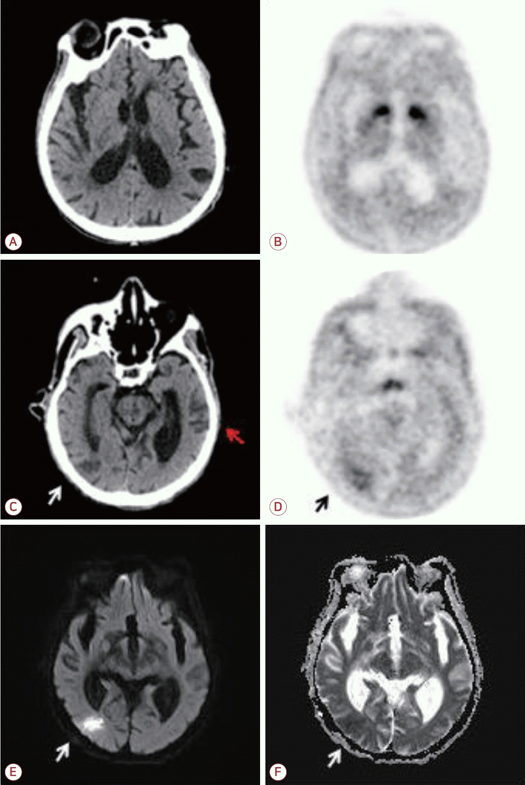
Figure.

Brain images of the patient. Compared to the brain CT, 18F-FP-CIT PET/CT demonstrated asymmetrically decreased FP-CIT uptake in the bilateral posterior putamen (A, B). Also, it revealed an unusual increased CIT uptake in 18F-FP-CIT PET/CT at same region where hypodense lesion is observed in the cortico-subcortical region of the right occipital lobe (C, D, white and black arrow). Diffusion-weighted MR images showed restricted diffusion with reduced apparent diffusion coefficient in the corresponding area (E, F, white arrow). In contrast, the hypodense lesion in the left temporal lobe does not show increased CIT uptake or diffusion restriction and is a finding of chronic infarction on brain CT (C, red arrow). CT; computed tomography, 18F-FP-CIT; 18F-fluorinated-N-3-fluoropropyl-2-b-carboxymethoxy-3-b-(4-iodophenyl) nortropane, PET; positron emission tomography, MR; magnetic resonance.

해당 부위는 뇌 computed tomography에서 저음영으로, 확산강조영상에서 확산 제한을 보여 급성 또는 아급성 뇌경색임을 확인하였다(Fig. C-F). 대조적으로 좌측 측두엽의 만성 뇌경색 병변에는 CIT 섭취 증가가 보이지 않았다(Fig. C, D). 이런 소견은 급성기에 뇌혈액장벽의 손상에 의해 CIT가 뇌실질로 누출되어 발생하였을 가능성과[1] 뇌경색 급성기 동안 활성화된 교세포와 골수 유래 대식세포에서 일시적으로 도파민D2수용체가 발현하여 보이는 것일 수 있다[2].

References

1. Cheon M, Lee KH. Focal increased 18F FP-CIT uptake in a recent ischemic lesion in the frontal lobe. Clin Nucl Med 2015;40:e137–e139.
2. Huck JH, Freyer D, Böttcher C, Mladinov M, Muselmann-Genschow C, Thielke M, et al. De novo expression of dopamine D2 receptors on microglia after stroke. J Cereb Blood Flow Metab 2015;35:1804–1811.

Article information Continued

Figure.

Brain images of the patient. Compared to the brain CT, 18F-FP-CIT PET/CT demonstrated asymmetrically decreased FP-CIT uptake in the bilateral posterior putamen (A, B). Also, it revealed an unusual increased CIT uptake in 18F-FP-CIT PET/CT at same region where hypodense lesion is observed in the cortico-subcortical region of the right occipital lobe (C, D, white and black arrow). Diffusion-weighted MR images showed restricted diffusion with reduced apparent diffusion coefficient in the corresponding area (E, F, white arrow). In contrast, the hypodense lesion in the left temporal lobe does not show increased CIT uptake or diffusion restriction and is a finding of chronic infarction on brain CT (C, red arrow). CT; computed tomography, 18F-FP-CIT; 18F-fluorinated-N-3-fluoropropyl-2-b-carboxymethoxy-3-b-(4-iodophenyl) nortropane, PET; positron emission tomography, MR; magnetic resonance.