피질척수로와 안 쪽 섬유띠의 변성을 보이는 성인 발병 크라베병

Adult-Onset Krabbe Disease with MRI Hyperintensities Along Corticospinal Tract and Medial Lemniscus

Article information

J Korean Neurol Assoc. 2022;40(1):85-86
Publication date (electronic) : February 1, 2022
doi : http://dx.doi.org/10.17340/jkna.2022.1.17
Department of Neurology, Ajou University School of Medicine, Suwon, Korea
박동규, 윤정한, 주인수
아주대학교 의과대학 신경과
Address for correspondence: Don Gueu Park, MD Department of Neurology, Ajou University School of Medicine, 164 World cup-ro, Yeongtong-gu, Suwon16499, Korea Tel: +82-31-219-7852 Fax: +82-31-219-5178 E-mail: dgpark17@gmail.com
received : August 11, 2021 , rev-recd : October 5, 2021 , accepted : October 5, 2021 .

65세 여자가 15년 전부터 서서히 진행하는 경직 하반신마비로 왔다. 양측 하지 근력이 Medical Research Council 4등급으로 저하되었고 건반사가 항진되었으며 바뱅스키징후 또한 관찰되었다. 근전도검사 상 특이 소견은 보이지 않았으나, 시행한 뇌 자기공명영상에서 양측 피질척수로 및 뇌교의 안 쪽 섬유띠를 포함하는 변성이 관찰되었다(Fig.).
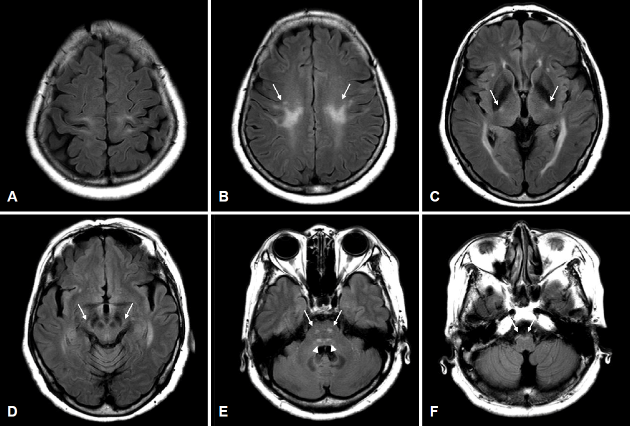
Figure.

Axial MRI of the brain with fluid-attenuated inversion recovery (FLAIR) sequence showing symmetric hyperintensities in (A) precentral gyri, (B) corona radiata, (C) posterior limbs of internal capsule, (D) cerebral peduncles of midbrain, (E) ventral pons, and (F) pyramids of medulla, which correspond to the degeneration of corticospinal tract (long arrows). Symmetric hyperintensities are also noted in (E) pontine tegmentum, corresponding to medial lemniscus (arrowheads). MRI; magnetic resonance imaging.

환자는 신경질환의 가족력은 없었으나 유전 경직 하반신마비 감별을 위하여 차세대 염기서열분석을 시행한 결과, 크라베(Krabbe)병과 연관된 GALC 유전자에서 기존에 알려진 병원성/유사병원성 변이가 발견되었다(c.[1901T>C];[683_649delinsCTC], p.[L634S]; [N228_S232delinsTP]) [1]. 크라베병은 상염색체열성으로 유전되는 드문 지질침착질환으로 주로 소아에서 발병하나 드물게는 성인에서도 발병한다. 피질척수로를 따라 나타나는 백질변성은 크라베병의 특징이며, 안 쪽 섬유띠 병변 또한 천막하 병변 중에서는 가장 흔하다고 알려져 있다[2]. 경직 하반신마비 환자에서 위와 같은 특징 적인 자기공명영상이 보일 경우 유전자검사를 고려하여야 하겠다.

References

1. Lim SM, Choi BO, Oh SI, Choi WJ, Oh KW, Nahm M, et al. Patient fibroblasts-derived induced neurons demonstrate autonomous neuronal defects in adult-onset Krabbe disease. Oncotarget 2016;7:74496–74509.
2. Cousyn L, Law-Ye B, Pyatigorskaya N, Debs R, Froissart R, Piraud M, et al. Brain MRI features and scoring of leukodystrophy in adult-onset Krabbe disease. Neurology 2019;93:e647–e652.

Article information Continued

Figure.

Axial MRI of the brain with fluid-attenuated inversion recovery (FLAIR) sequence showing symmetric hyperintensities in (A) precentral gyri, (B) corona radiata, (C) posterior limbs of internal capsule, (D) cerebral peduncles of midbrain, (E) ventral pons, and (F) pyramids of medulla, which correspond to the degeneration of corticospinal tract (long arrows). Symmetric hyperintensities are also noted in (E) pontine tegmentum, corresponding to medial lemniscus (arrowheads). MRI; magnetic resonance imaging.