척수압박을 유발한 IgG4-연관척수경수막염
IgG4-related Spinal Pachymeningitis Causing Spinal Cord Compression
Article information
64세 여자가 사지 근력 저하 및 경부 통증으로 병원에 왔다. 4년 전 두통으로 뇌자기공명영상에서 큰구멍(foramen magnum) 주변의 종양을 진단받고 절제수술을 받은 과거력이 있었다. 척수 자기공명영상에서 C1에서 T4까지 이어지는 경막외척수압박증이 확인되어(Fig.) 감압수술 및 개방 생검을 진행하였다. 병리조직 검사 결과 림프구형질세포 과다 침윤이 확인되었고 면역조직화학염색에서 많은 immunoglobulin G4 (IgG4) 양성 형질세포가 확인되어 최종적으로 IgG4-연관척수경수막염으로 진단되었다.
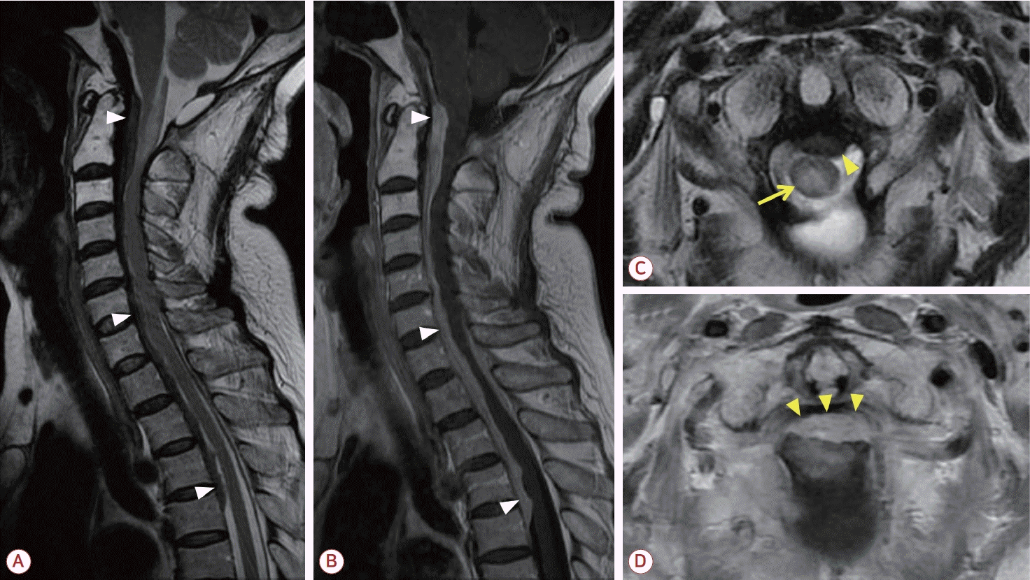
(A) Sagittal T2-weighted, (B) sagittal gadolinium-enhanced T1-weighted magnetic resonance imaging (MRI) showed an elongated spindle-shaped dural mass lesion over the anterior and posterior borders of the spinal cord at the C1-T4 levels (white arrowheads). (C) Axial T2-weighted MRI showed a dural mass lesion (yellow arrowhead) and intramedullary high-signal intensity lesion (yellow arrow) of the spinal cord at C1/2 level. (D) Axial gadolinium-enhanced T1-weighted MRI showed a dural mass lesion causing spinal cord compression at C1/2 level (yellow arrowheads).
IgG4-연관질환은 비교적 최근에 알려지기 시작한 전신 섬유염증질환으로, 신경계에서는 비후경수막염, 뇌하수체 침범, 안와질환이 주로 보고되었다[1]. IgG4-연관척수경수막염의 경우 영상 소견만으로는 수막종, 전이 종양 등으로 오인할 수 있고[2], 감압수술과 더불어 면역 치료까지 요하는 경우가 많으므로 감별해야 할 질환으로 포함되어야 한다.