

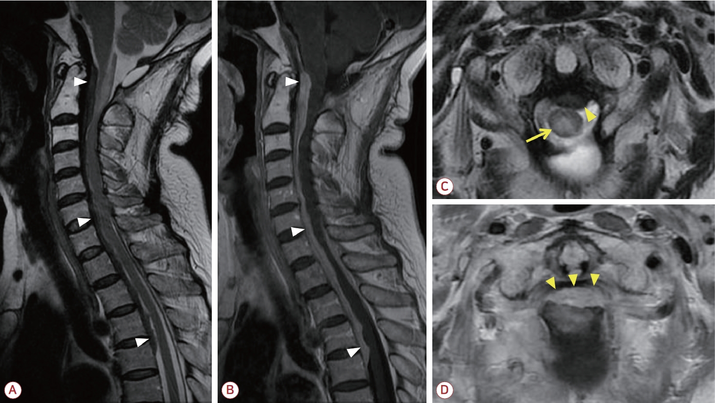
64세 여자가 사지 근력 저하 및 경부 통증으로 병원에 왔다. 4년 전 두통으로 뇌자기공명영상에서 큰구멍(foramen magnum) 주변의 종양을 진단받고 절제수술을 받은 과거력이 있었다. 척수 자기공명영상에서 C1에서 T4까지 이어지는 경막외척수압박증이 확인되어(Fig.) 감압수술 및 개방 생검을 진행하였다. 병리조직 검사 결과 림프구형질세포 과다 침윤이 확인되었고 면역조직화학염색에서 많은 immunoglobulin G4 (IgG4) 양성 형질세포가 확인되어 최종적으로 IgG4-연관척수경수막염으로 진단되었다.
| J Korean Neurol Assoc > Volume 42(1); 2024 > Article |

|
REFERENCES
1. AbdelRazek MA, Venna N, Stone JH. IgG4-related disease of the central and peripheral nervous systems. Lancet Neurol 2018;17:183-192.


2. Sbeih I, Darwazeh R, Shehadeh M, Al-Kanash R, Abu-Farsakh H, Sbeih A. Immunoglobulin G4-related hypertrophic pachymeningitis of the spine: a case report and systematic review of the literature. World Neurosurg 2020;143:445-453.
Figure.
(A) Sagittal T2-weighted, (B) sagittal gadolinium-enhanced T1-weighted magnetic resonance imaging (MRI) showed an elongated spindle-shaped dural mass lesion over the anterior and posterior borders of the spinal cord at the C1-T4 levels (white arrowheads). (C) Axial T2-weighted MRI showed a dural mass lesion (yellow arrowhead) and intramedullary high-signal intensity lesion (yellow arrow) of the spinal cord at C1/2 level. (D) Axial gadolinium-enhanced T1-weighted MRI showed a dural mass lesion causing spinal cord compression at C1/2 level (yellow arrowheads).
- TOOLS
 PDF Links
 PubReader
 ePub Link
 Full text via DOI
 Download Citation
 Print
Print


-
METRICS

-
- 0 Crossref
- 0 Scopus
- 533 View
- 26 Download
-

- Related articles
-
IgG4-Related Hypertrophic Pachymeningitis Mimicking Cerebral Venous Thrombosis2018 August;36(3)
- Editorial Office
-
(ZIP 03163) #1111, Daeil Bldg, 12, Insadong-gil, Jongno-gu, Seoul, Korea
Tel: +82-2-737-6530 Fax: +82-2-737-6531 E-mail: jkna@neuro.or.kr
Copyright © 2024 by Korean Neurological Association.

