경동맥갈퀴막 진단에 있어서 경동맥이중초음파검사의 유용성
Usefulness of Carotid Duplex Ultrasonography for Carotid Web
Article information
고혈압과 비대심근병증으로 약물 복용 중인 90세 여자 환자가 언어장애로 내원하였다. 신경계진찰에서 전실어증과 좌측으로 안구 편위, 우측 편마비가 확인되었다. 뇌 자기공명영상촬영에서 좌측 원위부 중간대뇌동맥 협착으로 인한 뇌경색이 확인되었다(Fig. A, B). 좌측경동맥에 얇은 선반모양의 구조물로 인한 충전결손(filling defect)이 의심되어 시행한 경동맥이중초음파검사에서 좌측경동맥 팽대부에 끝으로 갈수록 얇아지는 고에코경화판(hyperechoic plaque)과 주변으로 혈류 정체가 확인되었다(Fig. C-E). 경동맥갈퀴막과 연관된 뇌경색으로 진단하여 항응고제 치료를 시작하였다.
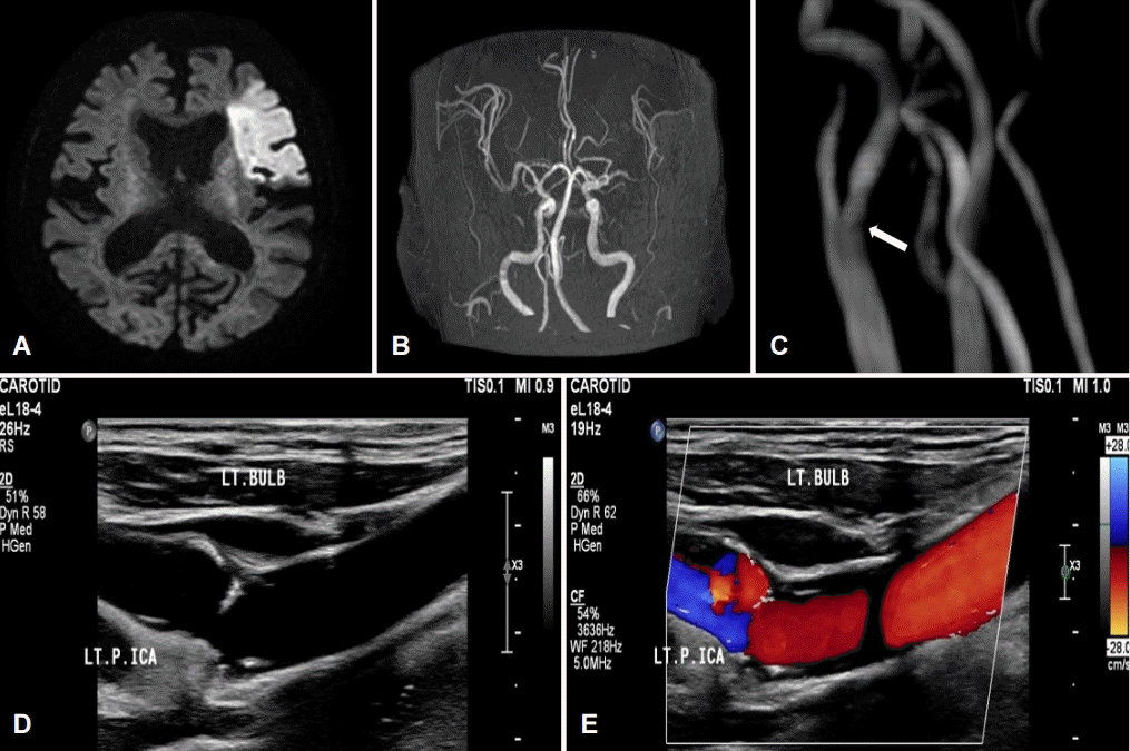
(A) Brain magnetic resonance diffusion-weighted image shows high signal intensity in the territory of the left MCA. (B, C) Brain magnetic resonance angiography shows occlusion of left distal MCA and suspicious shelf-like filling defect of left ICA (arrow). (D) Carotid duplex ultrasonography shows hyperechoic shelf-like plaque and (E) color Doppler shows stasis of blood flow in left ICA. LT; left, MCA; middle cerebral artery, ICA; internal cerebral artery.
경동맥갈퀴막에서 와류(turbulent flow)와 혈류의 정체(stasis)는 혈전을 만들게 되고 이는 색전성 뇌경색을 유발하므로 빠른 진단이 필요하다[1,2]. 디지털감산혈관조영술을 통하여 정확한 진단을 하게 되지만 침습적인 시술이 힘든 고령의 환자에서는 경동맥이중초음파검사를 통하여 갈퀴막과 주변 혈류 이상을 확인한다면 치료 방법을 결정하는 데 도움이 될 수 있다[1,2].