

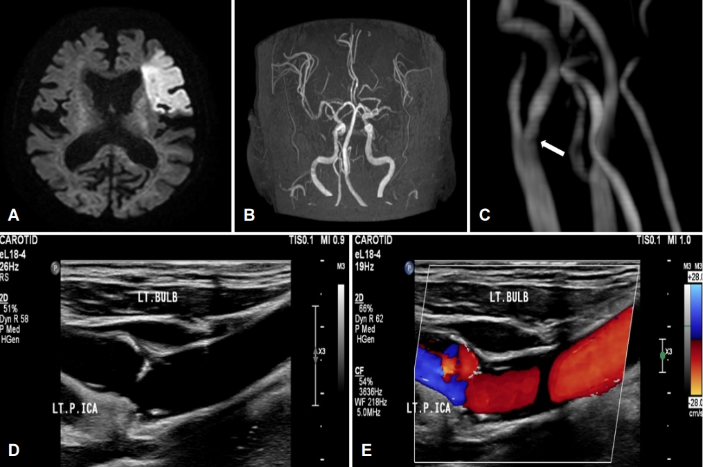
고혈압과 비대심근병증으로 약물 복용 중인 90세 여자 환자가 언어장애로 내원하였다. 신경계진찰에서 전실어증과 좌측으로 안구 편위, 우측 편마비가 확인되었다. 뇌 자기공명영상촬영에서 좌측 원위부 중간대뇌동맥 협착으로 인한 뇌경색이 확인되었다(Fig. A, B). 좌측경동맥에 얇은 선반모양의 구조물로 인한 충전결손(filling defect)이 의심되어 시행한 경동맥이중초음파검사에서 좌측경동맥 팽대부에 끝으로 갈수록 얇아지는 고에코경화판(hyperechoic plaque)과 주변으로 혈류 정체가 확인되었다(Fig. C-E). 경동맥갈퀴막과 연관된 뇌경색으로 진단하여 항응고제 치료를 시작하였다.
| J Korean Neurol Assoc > Volume 40(3); 2022 > Article |

|
REFERENCES
1. Zhang AJ, Dhruv P, Choi P, Bakker C, Koffel J, Anderson D, et al. A systematic literature review of patients with carotid web and acute ischemic stroke. Stroke 2018;49:2872-2876.


2. Park HK, Hong KS. Carotid web:under-recognized etiology for ischemic stroke. J Neurosonol Neuroimag 2018;10:100-105.
Figure.
(A) Brain magnetic resonance diffusion-weighted image shows high signal intensity in the territory of the left MCA. (B, C) Brain magnetic resonance angiography shows occlusion of left distal MCA and suspicious shelf-like filling defect of left ICA (arrow). (D) Carotid duplex ultrasonography shows hyperechoic shelf-like plaque and (E) color Doppler shows stasis of blood flow in left ICA. LT; left, MCA; middle cerebral artery, ICA; internal cerebral artery.
- TOOLS
 PDF Links
 PubReader
 ePub Link
 Full text via DOI
 Download Citation
 Print
Print


-
METRICS

-
- 0 Crossref
- 0 Scopus
- 794 View
- 57 Download
-

- Related articles
-
Usefulness of Transcranial Doppler Sonography for Determining Brain Death2015 May;33(2)
Hypoplasia of the Internal Carotid Artery: Duplex Ultrasonographic Findings2012 ;30(1)
- Editorial Office
-
(ZIP 03163) #1111, Daeil Bldg, 12, Insadong-gil, Jongno-gu, Seoul, Korea
Tel: +82-2-737-6530 Fax: +82-2-737-6531 E-mail: jkna@neuro.or.kr
Copyright © 2024 by Korean Neurological Association.

