무증상 대동맥박리 진단을 위한 목동맥초음파에서 관찰된 온목동맥박리의 유용성
Clinical Usefulness of Common Carotid Artery Dissection in Carotid Ultrasound for the Diagnosis of Asymptomatic Aortic Dissection
Article information
63세 여자가 혼미, 좌측 반신마비(Medical Research Council grade I)로 병원에 왔다. National Institute of Health (NIH) 뇌졸중 척도 19점으로 computed tomography (CT) 촬영 후 정맥내혈전용해제(recombinant tissue plasminogen activator)를 사용하였다. CT혈관조영술 및 뇌 자기공명영상(magnetic resonance imaging)에서 우측 중대뇌동맥영역의 뇌경색과 우측 중대뇌동맥 M2-분절의 협착이 관찰되었다(Fig. 1A-C). 혈전용해제 투여 4시간 후 환자는 NIH 뇌졸중척도 3점으로 호전을 보였다. 이중목동맥초음파에서 우측 온목동맥의 내막판과 거짓속공간이 관찰되어 동맥박리를 진단하였고(Fig. 1-D, E), 함께 시행한 대동맥 CT에서 오름대동맥에서 엉덩동맥갈림까지의 동맥박리를 확인하였다(Fig. 2).
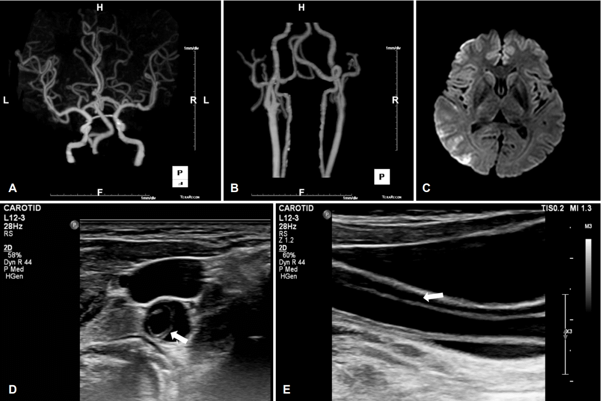
Neuroimages of the patient. (A, B) Carotid CT angiography reveals right MCA M2 stenosis. (C) Diffusion weighted magnetic resonance images shows multiple acute cerebral infarction at right MCA territory. Transverse (D) and longitudinal (E) B-mode images of right CCA show movable intimal flap (arrow) and false lumen. CT; computed tomography, MCA; middle cerebral artery, CCA, common carotid artery.
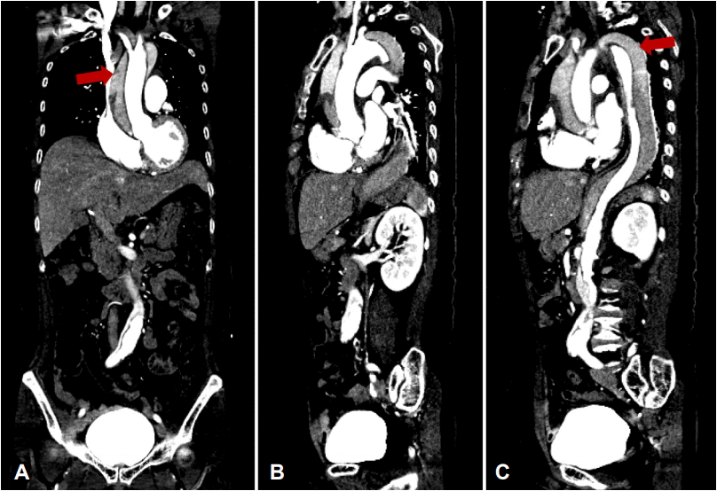
(A-C) Aorto-abdominal vascular CT image show aortic dissection (red arrows) in ascending, aortic arch, and descending aorta (Stanford type A, Debakey type I). CT; computed tomography.
무증상 대동맥박리는 전형적인 흉통 등의 증상 없이 신경계증상만 관찰된 경우 진단이 지연될 수 있고, 이를 인지하지 못하고 혈전용해제 등의 치료를 할 경우 심장눌림증이나 혈흉 등의 심각한 합병증을 유발할 수 있다[1]. 목동맥초음파는 진단 특이도가 높은 비침습검사로 빠르게 광범위한 동맥박리를 확인할 수 있으며[2], 특히 온목동맥박리가 확인되는 경우 동반되는 대동맥혈관박리에 대한 추가적인 혈관 평가가 필요하다.