

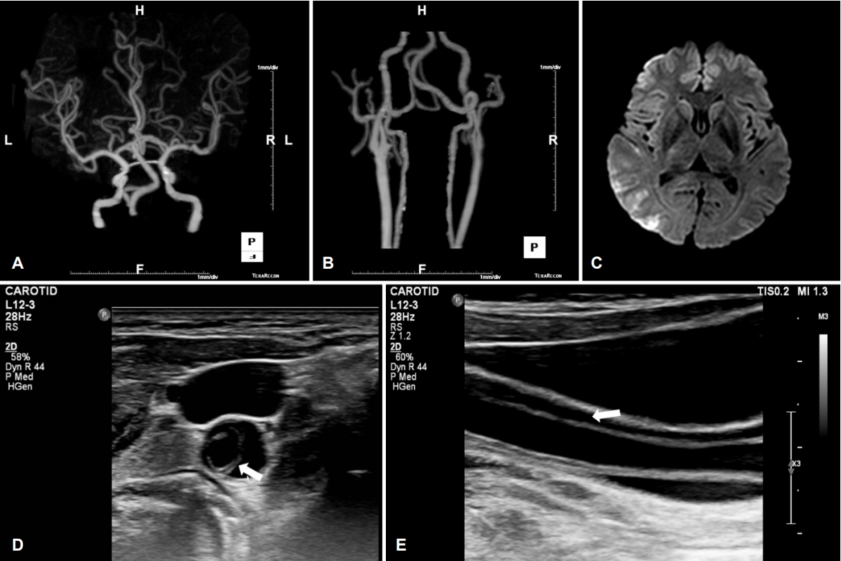
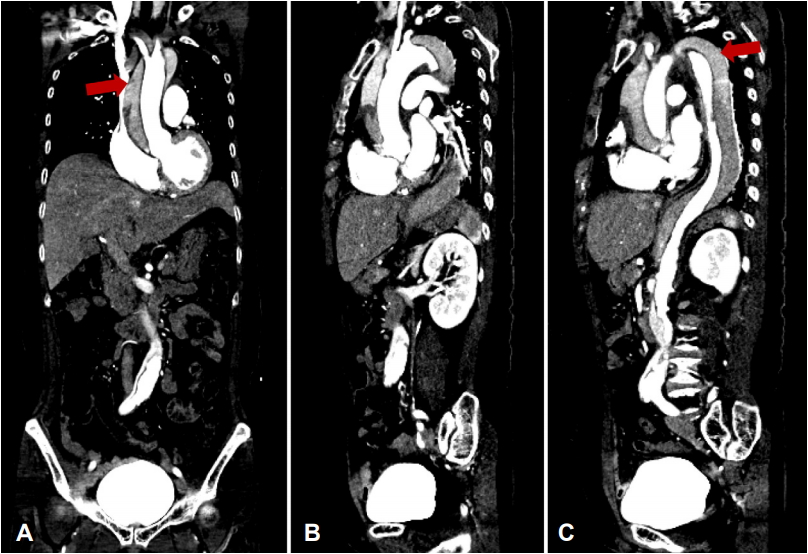
63세 여자가 혼미, 좌측 반신마비(Medical Research Council grade I)로 병원에 왔다. National Institute of Health (NIH) 뇌졸중 척도 19점으로 computed tomography (CT) 촬영 후 정맥내혈전용해제(recombinant tissue plasminogen activator)를 사용하였다. CT혈관조영술 및 뇌 자기공명영상(magnetic resonance imaging)에서 우측 중대뇌동맥영역의 뇌경색과 우측 중대뇌동맥 M2-분절의 협착이 관찰되었다(Fig. 1A-C). 혈전용해제 투여 4시간 후 환자는 NIH 뇌졸중척도 3점으로 호전을 보였다. 이중목동맥초음파에서 우측 온목동맥의 내막판과 거짓속공간이 관찰되어 동맥박리를 진단하였고(Fig. 1-D, E), 함께 시행한 대동맥 CT에서 오름대동맥에서 엉덩동맥갈림까지의 동맥박리를 확인하였다(Fig. 2).
| J Korean Neurol Assoc > Volume 38(1); 2020 > Article |

|
Figure 1.
Neuroimages of the patient. (A, B) Carotid CT angiography reveals right MCA M2 stenosis. (C) Diffusion weighted magnetic resonance images shows multiple acute cerebral infarction at right MCA territory. Transverse (D) and longitudinal (E) B-mode images of right CCA show movable intimal flap (arrow) and false lumen. CT; computed tomography, MCA; middle cerebral artery, CCA, common carotid artery.
- TOOLS
 PDF Links
 PubReader
 ePub Link
 Full text via DOI
 Download Citation
 Print
Print


-
METRICS

-
- 0 Crossref
- 0 Scopus
- 4,682 View
- 90 Download
- Related articles
- Editorial Office
-
(ZIP 03163) #1111, Daeil Bldg, 12, Insadong-gil, Jongno-gu, Seoul, Korea
Tel: +82-2-737-6530 Fax: +82-2-737-6531 E-mail: jkna@neuro.or.kr
Copyright © 2026 by Korean Neurological Association.

