갑상샘눈병증 관련 하직근 단독 비대

Isolated Enlargement of the Inferior Rectus Muscle Associated with Thyroid Eye Disease

Article information

J Korean Neurol Assoc. 2026;44(4):373-374
Publication date (electronic) : November 1, 2026
doi : http://dx.doi.org/10.17340/jkna.2026.0041
Department of Neurology, Veterans Health Service Medical Center, Seoul, Korea
윤보혜, 이슬, 김정섭, 이준수, 김승민
중앙보훈병원 신경과
Address for correspondence Seung Min Kim, MD, PhD Department of Neurology, Veterans Health Service Medical Center, 53 Jinhwangdo-ro 61-gil, Gangdong-gu, Seoul 05368, Korea Tel: +82-2-2225-1324 Fax: +82-2-2225-1327 E-mail: kh21762@hanmail.net
received : June 9, 2026 , rev-recd : July 16, 2026 , accepted : July 16, 2026 .

76세 남자가 한 달간 지속되는 양안 복시로 내원하였다. 아홉방향주시 검사에서 좌안 상전 및 외전 제한이 관찰되었고(Fig. A), 상방주시 시 안와 통증이 악화되었다. 동공반사, 시력, 시야는 정상이었고 다른 뇌신경학적 이상은 없었으나 단일 뇌신경마비로 설명하기 어려운 통증성 안구운동 제한을 보였다. 안와/외안근 질환, 복합 뇌신경마비, 신경근접합부 질환 감별을 위해 안와자기공명영상을 시행하였고 좌측 하직근의 국소 비대와 조영증강을 확인하였다(Fig. B, C). 갑상샘자극호르몬은 0.005 μIU/mL 미만으로 억제되었으나 티록신, 삼요오드티록신, 갑상샘자극호르몬수용체항체는 정상이어서 영상 소견까지 종합하여 무증상 갑상샘항진증 동반 갑상샘눈병증으로 판단하였다. 무증상 갑상샘항진증에서도 외안근 단독 비대가 가능하며[1], 하직근 비대에 의한 제한성 상전장애는 상직근 마비처럼 보일 수 있다. 이는 신경마비가 아닌 길항근의 기계적 제한이므로 안와자기공명영상의 외안근 평가가 진단에 중요하다[2].
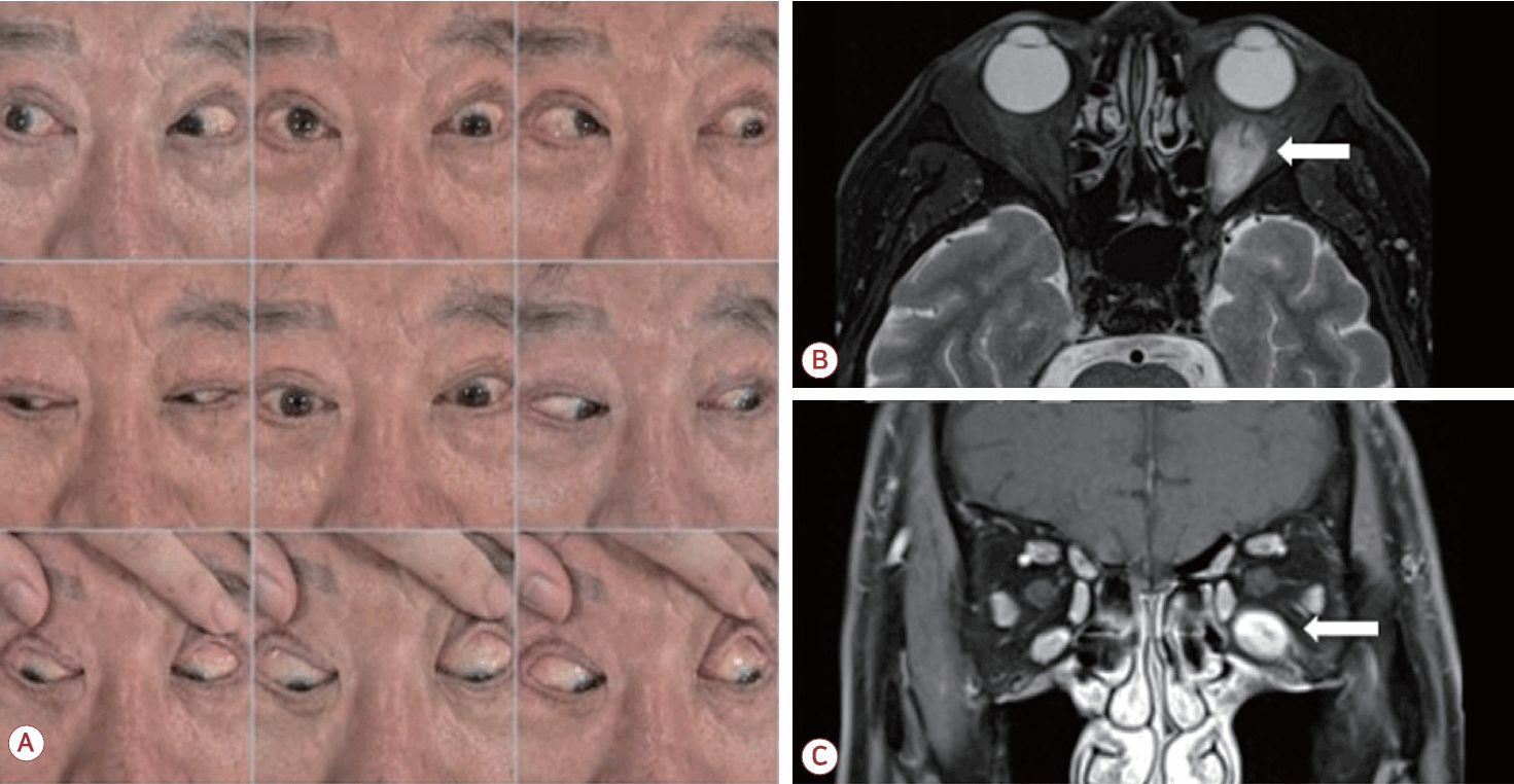
Figure.

(A) Nine-gaze photographs demonstrate impaired elevation and abduction of the left eye. (B) Axial T2-weighted orbital magnetic resonance image and (C) contrast-enhanced coronal T1-weighted image demonstrate isolated enlargement and enhancement of the left inferior rectus muscle (arrow).

References

1. Bahn RS. Graves’ ophthalmopathy. N Engl J Med 2010;362:726–738.
2. Wiersinga WM, Eckstein AK, Žarković M. Thyroid eye disease (Graves’ orbitopathy): clinical presentation, epidemiology, pathogenesis, and management. Lancet Diabetes Endocrinol 2025;13:600–614.

Article information Continued

Figure.

(A) Nine-gaze photographs demonstrate impaired elevation and abduction of the left eye. (B) Axial T2-weighted orbital magnetic resonance image and (C) contrast-enhanced coronal T1-weighted image demonstrate isolated enlargement and enhancement of the left inferior rectus muscle (arrow).