자기공명영상조영술에서 M1 협착으로 의심된 중대뇌동맥 Fenestration
Middle Cerebral Artery Fenestration Mimicking Proximal M1 Stenosis on Time-of-Flight Magnetic Resonance Angiography
Article information
A 65-year-old woman with sudden-onset headache was referred after time-of-flight magnetic resonance angiography suggested severe stenosis of the proximal right M1 segment (Fig. A). Computed tomography angiography at our institution demonstrated M1 fenestration with two parallel channels rejoining distally, considered incidental to the headache (Fig. B, C). Middle cerebral artery fenestration is rare, with a reported prevalence of approximately 0.15% [1]. Local flow disturbance and thrombus formation have been proposed as potential mechanisms of ischemia [2]. Poor visualization of one channel may mimic focal stenosis, whereas a double-lumen appearance may resemble dissection [3]. Atypical or clinically discordant M1 narrowing warrants consideration of fenestration and further vascular imaging.
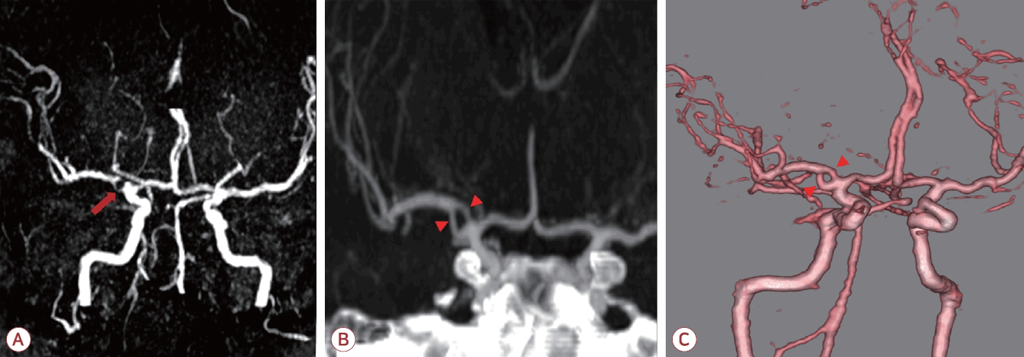
(A) Time-of-flight magnetic resonance angiography obtained at the referring hospital shows apparent severe focal stenosis of the proximal right M1 segment (arrow). (B) Magnified maximum-intensity projection and (C) three-dimensional volume-rendered computed tomography angiography obtained at our institution demonstrate a short-segment M1 fenestration, with the artery dividing into two parallel channels and rejoining distally (arrowheads).
Notes
ACKNOWLEDGEMENTS
This work was supported by Wonkwang University in 2026.