진행성 다초점 백질뇌병증의 자화율 강조 영상에서 관찰되는 운동 띠 징후 유사 소견
Motor Band Sign-Like Appearance on Susceptibility-Weighted Imaging in Possible Progressive Multifocal Leukoencephalopathy
Article information
The “motor band sign (MBS)” is a loss of signal in the precentral gyrus on susceptibility-weighted imaging (SWI), which is known to result from iron accumulation in the motor cortex. This finding has emerged as a useful radiological marker of amyotrophic lateral sclerosis (ALS), correlating with upper motor neuron impairment [1,2]. However, the MBS is not entirely specific to ALS, and has been reported in other neurological conditions including cerebral infarction, Parkinson’s disease, and central nervous system (CNS) infectious or inflammatory disorders [3]. Progressive multifocal leukoencephalopathy (PML), caused by JC virus (JCV) reactivation in immunocompromised hosts, can show subcortical white matter U-fiber involvement and cortical lesions with low signal intensity rim on SWI [4]; localized PML involving the motor cortex may radiologically mimic the MBS. We report a case of possible PML in an human immunodeficiency virus (HIV) patient whose initial presentation and SWI findings closely mimicked the MBS of ALS.
CASE
A 68-year-old man presented with a 5-month history of progressive left-hand weakness. He had been diagnosed with HIV infection 12 years prior to presentation, with an initial CD4 count of 13.2 cells/μL but had been lost to follow-up for the preceding 9 years. Neurological examination showed isolated left upper extremity weakness with a distal predominance (Medical Research Council grade 2-3 distally and 4+ proximally). Sensory examination was intact and mild hyperreflexia was noted in the affected limb. Electrophysiological studies showed only mild left ulnar motor nerve slowing at the elbow without evidence of active denervation, which was insufficient to explain the profound clinical weakness.
Brain magnetic resonance imaging (MRI) showed a non-enhancing lesion in the right frontoparietal white matter on fluid-attenuated inversion recovery (FLAIR) images (Fig. B). Notably, SWI sequences revealed a distinct linear hypointensity along the right precentral gyrus (Fig. A), strikingly similar to the MBS typically associated with ALS [5]. Cerebrospinal fluid (CSF) evaluation was unremarkable and negative for common opportunistic infections. Although CSF JCV DNA polymerase chain reaction (PCR) was negative, the patient was diagnosed with possible PML according to the American Academy of Neurology (AAN) criteria, based on severe immunosuppression (CD4 count of 3.8 cells/μL, HIV RNA load of 37,300 copies/mL), the characteristic MRI findings, and a compatible clinical features. Antiretroviral therapy was therefore immediately re-initiated. On follow-up MRI after 6 months later, the previously non-enhancing lesion had become slightly edematous with new contrast enhancement, findings suggestive of PML-associated immune reconstitution inflammatory syndrome (PML-IRIS) and further supporting the diagnosis. Cortical hypointensity on SWI persisted (Fig. C), accompanied by localized cortical atrophy and adjacent white matter abnormality on FLAIR (Fig. D), possibly reflecting tissue destruction associated with the PML process.
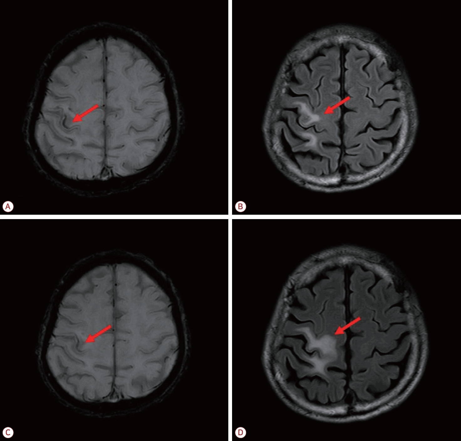
Brain MRI at presentation and at 6-month follow-up. (A, B) Initial susceptibility-weighted imaging (SWI) and fluid-attenuated inversion recovery (FLAIR) images demonstrate a unilateral motor band sign-like cortical hypointensity in the right precentral gyrus (A, arrow) that spatially corresponds to the white matter lesion of progressive multifocal leukoencephalopathy (PML) (B, arrow). (C, D) Follow-up SWI and FLAIR images obtained at 6-month later show persistent cortical hypointensity (C, arrow) together with progression of the adjacent white matter abnormality and localized cortical atrophy (D, arrow), possibly reflecting ongoing tissue destruction by PML.
CONCLUSION
Although both ALS and PML can produce linear SWI hypointensity along the precentral gyrus, the underlying pathomechanisms might differ. In ALS, the MBS reflects iron accumulation within the microglia secondary to upper motor neuron degeneration [1]. In PML, cortical SWI hypointensity is thought to arise from a cascade of JCV-mediated demyelination and late-stage neuroinflammation, ultimately leading to axonal loss and subsequent hemosiderin deposition [6,7]. Additionally, the MBS in ALS typically appears bilaterally [2,5], while PML usually produces asymmetric brain lesions. Nevertheless, this case demonstrates that localized PML restricted to the motor cortex can produce SWI findings similar to the MBS, particularly in patients presenting with slowly progressive monoparesis. A limitation of this report is that the CSF JCV PCR was negative and histopathological confirmation was not obtained, classifying this case as possible PML. In patients with advanced HIV presenting with focal white matter lesions, alternative etiologies such as primary CNS lymphoma or other opportunistic infections must be carefully considered. However, these were effectively excluded through unremarkable laboratory results. Furthermore, the highly characteristic neuroimaging features, combined with the clinical course with subsequent development of IRIS during treatment, strongly support the diagnosis of PML. Clinicians should be aware that MBS-like SWI findings are not pathognomonic for ALS and that focal destructive processes including PML can produce a similar radiological appearance.