COL4A2 c.941del 변이를 동반한 양측 중대뇌동맥 혈관병증 환자에서 발생한 좌측 중대뇌동맥 관통동맥 영역 급성 뇌경색
Acute Left Middle Cerebral Artery Perforator Territory Infarction in a Patient with Bilateral Middle Cerebral Artery Vasculopathy and a Rare COL4A2 c.941del Variant
Article information
Trans Abstract
We report a 25-year-old man with acute left middle cerebral artery perforator territory infarction, bilateral middle cerebral artery stenoses, right M1 vessel-wall enhancement suggesting non-atherosclerotic vasculopathy, and a rare heterozygous COL4A2 c.941del frameshift variant, highlighting the need to consider COL4A1/A2-related vasculopathy in young stroke patients with atypical bilateral intracranial arterial abnormalities.
젊은 뇌졸중 환자에서 두개내동맥협착은 죽상경화뿐 아니라 모야모야병, 동맥 박리, 혈관염 등 다양한 원인으로 발생하며, 아시아인에서는 두개내죽상경화의 비중이 높다[1-3]. 그러나 젊고 전통적 위험인자가 적은 환자에서는 비죽상경화혈관병증이 죽상경화협착으로 오인될 수 있다. 최근 한국인 조기 발병 허혈뇌졸중 코호트에서 단일 유전자 뇌졸중 관련 변이가 드물지 않게 확인되었고[4] COL4A2는 IV형 콜라겐 α2 사슬을 부호화하여 혈관 기저막 안정성에 관여하며 다양한 뇌혈관 및 전신 표현형과 관련된다[5]. 저자들은 양측 중대뇌동맥 혈관병증과 좌측 중대 뇌동맥 관통동맥 영역 급성 뇌경색이 동반된 25세 남자에서 드문 COL4A2 c.941del 변이를 확인하였기에 이를 보고하고자 한다.
증 례
25세 남자가 내원 당일 발생한 우측 편마비와 감각 저하로 내원하였다. 우측 하지 grade IV+ 위약과 우측 반신 감각 저하(건측의 40%)가 있었다. 신장 178 cm, 체중 88 kg (체질량지수, 27.8)의 비흡연자였고 가족력은 없었다. 혈액 검사에서 저밀도 지단백질-콜레스테롤(Low-density Lipoprotein [LDL]-cholesterol) 192 mg/dL, 총 콜레스테롤 273 mg/dL, 중성지방 298 mg/dL의 이상지질혈증과 일시적 간효소 상승(아스파트산아미노기전달효소[Aspartate Aminotransferase, AST] 110 U/L, 알라닌아미노기전달효소[Alanine Aminotransferase, ALT] 270 U/L)이 있었으나 1개월 후 AST 42 U/L, ALT 81 U/L, 3개월 후 AST 31 U/L, ALT 50 U/L로 호전되었다. 신기능 검사, 요검사에 특이 소견은 없었고 심전도는 정상동 리듬이었으며 추적 심장 평가에서 난원공개존이 확인되어 경피적 폐색술을 시행하였다. 항인지질항체, C/S단백질, 호모시스테인, 항핵항체, 항중성구세포질항체, 류마티스인자, 한랭글로불린(cryoglobulin) 등 혈전 질환 및 혈관염 관련 검사에서 특이 소견은 없었다. 안과 검사는 시행하지 않았다. 뇌자기공명영상(Magnetic Resonance Imaging, MRI)에서 좌측 기저핵-내포 부위의 급성 뇌경색과 우측 중대뇌동맥 M1의 중등도-중증 협착, 좌측 M2 근위부 경도 협착이 확인되었다(Fig. 1). 3T 고해상혈관벽영상에서 우측 M1에 선형 혈관벽 조영증강, 양성자밀도강조 혈관벽영상에서 혈관벽내고신호가 관찰되었으나 이중내강(double lumen), 내막피판(intimal flap), 명확한 혈관벽내혈종(intramural hematoma)은 보이지 않았고 디지털감산혈관조영(digital subtraction angiography)은 시행되지 않았다. 이에 우측 M1 병변은 확정적 동맥박리보다 박리를 포함한 비죽상경화두개내혈관병증 가능성을 시사하는 혈관벽 이상 소견으로 판단하였다(Fig. 2). 표적 유전자 검사에서 COL4A2 NM_001846.4:c.941del(p.Gly314AspfsTer44) 이형 접합 변이가 확인되었고, 확인 가능한 인구 집단 데이터베이스에서 관찰되지 않았다. 검사 기관은 이 변이를 기능 손실(loss-of-function)이 질병 기전으로 알려진 유전자에서의 틀이동기능상실변이(frameshift null variant)로 해석하여 병원성의심변이(likely pathogenic variant)로 분류하였다. 해당 패널에는 COL4A1, NOTCH3, HTRA1, RNF213, ACTA2 , COL3A1, FBN1 등이 포함되었으나 COL4A2 c.941del 외에 다른 변이는 보고되지 않았다. 환자는 이중항혈소판제와 스타틴 치료 후 호전되어 퇴원하였다.
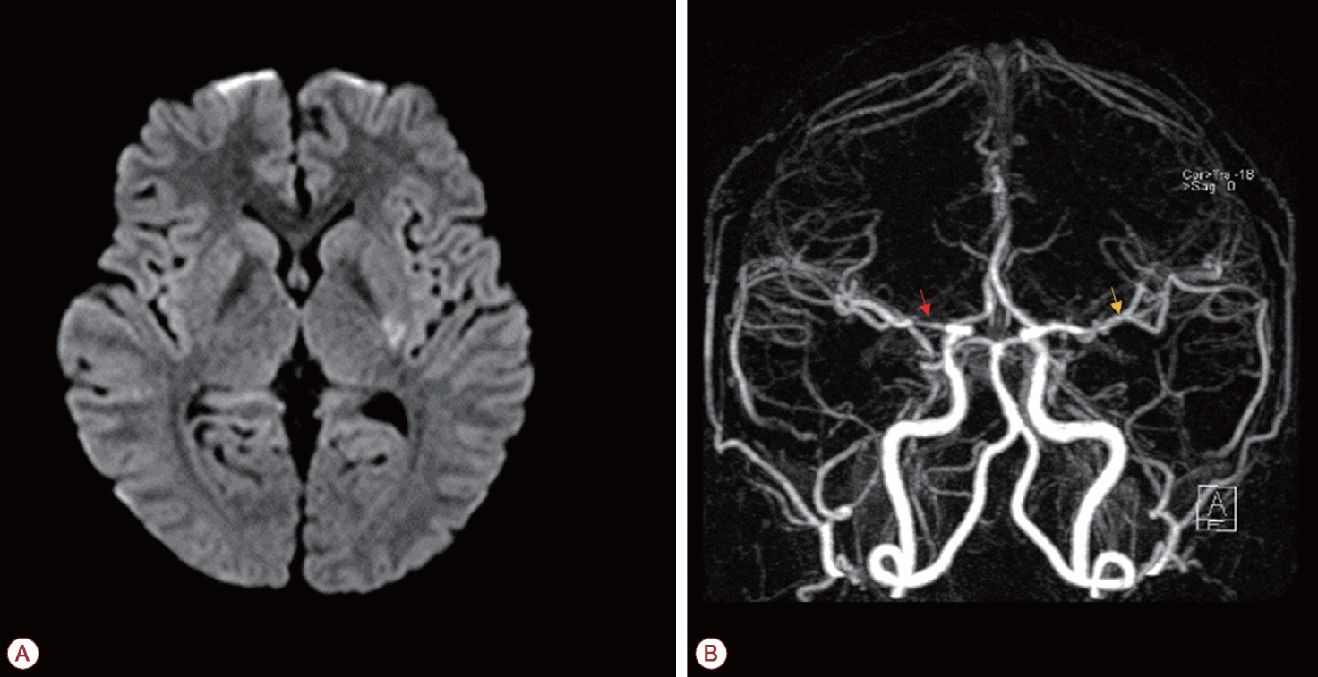
(A) Diffusion-weighted imaging shows an acute infarction in the left basal ganglia-internal capsule region, corresponding to the left middle cerebral artery (MCA) perforator territory. (B) MR angiography demonstrates moderate-to-severe segmental stenosis at the right M1 segment of the MCA (red arrow) and mild focal stenosis at the proximal left M2 segment (yellow arrow). MR; magnetic resonance.
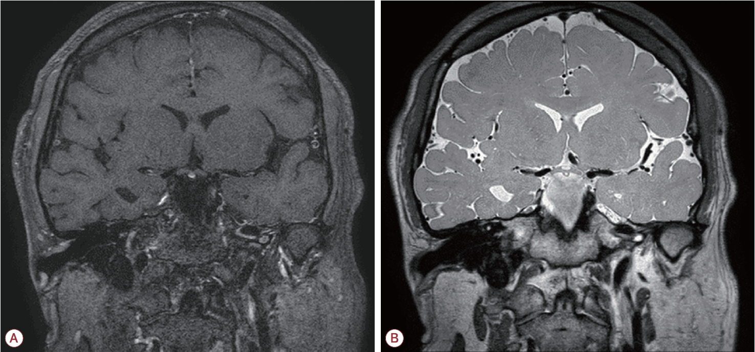
Three-tesla intracranial high-resolution vessel wall imaging of the right M1 segment of the MCA shows segmental stenosis with linear strong wall enhancement on contrast-enhanced T1-weighted images (A) and mild linear hyperintensity within the vessel wall on three-dimensional proton density-weighted vessel-wall images (B). These findings were interpreted as vessel-wall abnormalities suggesting non-atherosclerotic vasculopathy; however, definite imaging signs of arterial dissection, such as a double lumen or intimal flap, were not identified. MCA; middle cerebral artery.
고 찰
본 증례의 급성 뇌경색은 우측 M1 병변과 직접적인 인과 관계가 있는 병변이라기보다 좌측 중대뇌동맥 관통동맥 영역의 급성 뇌경색으로 해석하는 것이 적절하다. 난원공개존도 확인되어 색전성 기전 가능성도 고려해야 하나 양측 중대뇌동맥 혈관병증과 우측 M1 혈관벽 이상 소견은 단일 심장성 색전 기전만으로 설명하기 어렵다. 다만 우측 M1 혈관벽조영증강은 동맥박리를 시사할 수 있으나 단독으로 특이적이지 않고 혈관염 또는 다른 급성 혈관병증에서도 관찰될 수 있으므로 확정적 박리로 단정하기 어렵다. 한국인 일반 인구에서도 COL4A2 병적 변이가 보고된 바 있으나[6] 한국인 젊은 허혈뇌졸중 환자에서 본 증례처럼 양측 중대뇌동맥 혈관병증이 동반된 보고는 제한적이라는 점에서 의의가 있다. COL4A2 변이는 뇌내출혈, 백질변성, 뇌소혈관질환, 동맥류, 허혈뇌졸중 등 뇌혈관 표현형과 안과, 신장, 근육 침범 등 전신 표현형을 동반할 수 있다. COL4A2 c.941del은 Gly-X-Y 삼중나선 도메인의 글리신 잔기를 파괴하는 틀이동(frameshift) 조기 종결을 유발할 것으로 예측된다. 검사 기관에서는 병원성의심변이(likely pathogenic variant)로 분류하였으나 본 증례의 경우 해당 변이의 병원성과 임상 표현형 사이의 인과 관계가 가족 분리 분석이나 기능 연구로 확인되지 않았으므로 해석에 주의가 필요하다[5]. 또한 본 검사는 표적 유전자의 부호화 엑손(coding exon)을 대상으로 하므로 일부 구조 변이, 심부 인트론 및 조절부위 변이는 검출되지 않았을 수 있다. 젊은 뇌졸중 환자에서 전통적 혈관 위험인자로 충분히 설명되지 않는 양측성 또는 비전형두개내혈관병증이 관찰될 경우 COL4A1/A2 관련 질환을 포함한 단일 유전자 혈관병증을 감별 진단으로 고려할 필요가 있다.