우측 중대뇌동맥 영역 뇌경색에서 편측무시양 증상으로 나타난 좌측 동측반맹
Left Homonymous Hemianopia Mimicking Spatial Neglect in Right Middle Cerebral Artery Territory Infarction
Article information
Trans Abstract
Homonymous hemianopia, typically associated with posterior cerebral artery (PCA) infarction, can occur in middle cerebral artery (MCA) infarction involving the optic radiations. We report a 53-year-old man with right MCA infarction. After dysarthria and left-sided weakness improved, he developed gait deviation and neglect-like behavior. He was diagnosed with left homonymous hemianopia by visual field testing and automated perimetry and follow-up imaging showed no new PCA infarction. This case highlights the importance of visual field examination in MCA infarction.
동측반맹(homonymous hemianopia)은 교차뒤시각경로(retrochiasmal visual pathway) 병변의 대표적인 증상으로, 임상적으로는 흔히 후대뇌동맥(posterior cerebral artery) 영역의 뇌경색과 연관된다[1,2]. 그러나 시각부챗살(optic radiation)은 측두엽과 두정엽을 넓게 주행하므로 중대뇌동맥(middle cerebral artery) 영역 병변에서도 시야 결손이 발생할 수 있다[2-4]. 중대뇌동맥 뇌경색에서는 근력 저하, 구음장애, 편측무시(spatial neglect) 등이 전면에 나타나 시야장애가 간과되기 쉽지만 시야장애는 보행, 운전, 직업 복귀 및 일상생활 독립성에 영향을 미치는 기능적 장애이다[5-7]. 저자들은 우측 중대뇌동맥 영역 뇌경색 환자에서 편측무시양 증상으로 관찰되었으나 대면 시야 검사와 자동 시야 검사를 통해 좌측 동측반맹으로 확인된 증례를 보고하고자 한다.
증 례
53세 남자가 기상 후 인지한 구음장애와 좌측 근력 저하로 내원하였다. 응급실 내원 당시 혈압은 144/77 mmHg, 맥박수는 82회/분이었다. 신경계 진찰에서 명확한 시야 결손이나 편측무시를 시사하는 소견은 확인되지 않았으며 좌측 얼굴 마비, 경미한 구음장애 및 좌측 상하지의 근력 저하(Medical Research Council grade 4+)가 확인되어 NIH뇌졸중척도(National Institutes of Health Stroke Scale)는 4점이었다. 뇌자기공명영상(brain magnetic resonance imaging)의 확산강조영상에서 우측 중대뇌동맥 영역의 급성 뇌경색이 확인되었으며 병변은 우측 뒤쪽 측두두정 피질-피질하 영역(posterior temporoparietal cortico-subcortical region)을 중심으로 가쪽 측두후두 접합부(lateral temporo-occipital junction)와 인접한 심부백질(deep white matter)까지 분포하였다(Fig. 1-A). 전형적인 새발톱고랑 중심 후대뇌동맥 영역 병변이나 안쪽 후두엽 피질의 명확한 침범은 관찰되지 않았다.
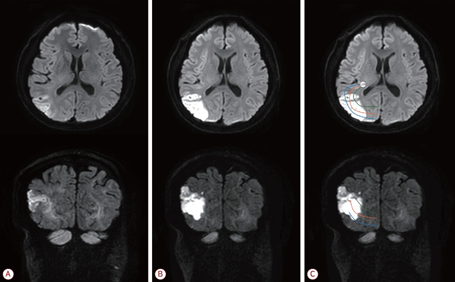
Serial diffusion-weighted magnetic resonance images and their relationship to the optic radiations. (A) Initial axial and coronal diffusion-weighted images show acute infarction in the right middle cerebral artery territory. (B) Follow-up axial and coronal diffusion- weighted images obtained after confirmation the visual field defect, show no newly developed acute infarction in the medial occipital cortex or typical posterior cerebral artery territory. (C) Schematic overlays on the follow-up images illustrate the estimated broad distribution and posterior convergence of the optic radiation bundles relative to the lesion. The blue, orange, and green lines represent the ventral bundle/Meyer’s loop, central bundle, and dorsal bundle of the optic radiations, respectively. The lateral geniculate nucleus (LGN) is marked in the axial image as an approximate anatomic landmark.
병일 2일째 구음장애와 좌측 근력 저하는 호전되어 보행이 가능하였으나 보행 중 좌측 공간을 충분히 인지하지 못하거나 의도한 방향과 다르게 이동하는 양상이 관찰되었다. 환자가 이러한 이상을 뚜렷하게 인지하지 못하여 처음에는 편측무시양 증상이 의심되었으나 대면 시야 검사에서 좌측 시야 결손이 의심되었다. 같은 날 시행한 신경심리 검사에서 레이복합도형 검사, 시계그리기 검사 및 글자지우기 검사를 시행하였으며 시공간 구성 능력 및 편측무시를 시사하는 이상 소견은 확인되지 않았다. 이어 시행한 자동 시야 검사에서 양안의 좌측 동측반맹이 확인되었다(Fig. 2).
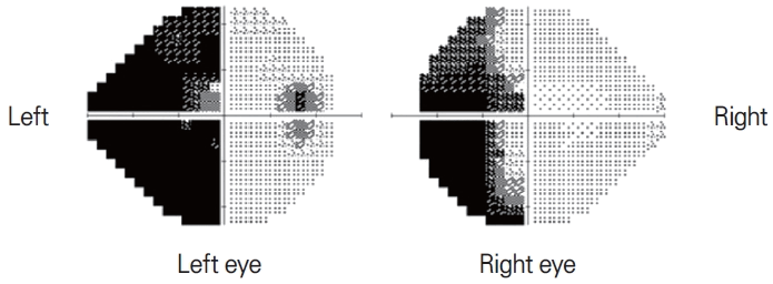
Automated perimetry. Automated perimetry demonstrates complete left homonymous hemianopia involving both the superior and inferior portions of the left visual hemifields of both eyes. The left and right labels represent the patient’s visual fields.
병일 5일째 시행한 추적 확산강조영상(follow-up diffusion-weighted imaging)에서도 기존 병변 외에 후두엽 또는 후대뇌동맥 영역의 새로운 급성 병변은 확인되지 않았다(Fig. 1-B). 환자는 급성기 치료 후 퇴원하였으며, 3개월 추적 관찰에서 주관적인 시야장애는 호소하지 않았고 대면 시야 검사에서 시야 결손이 확인되지 않았다. 임상적으로 시야장애가 호전된 것으로 판단되어 추적 자동 시야 검사는 시행하지 않았다.
고 찰
본 증례는 우측 중대뇌동맥 영역 뇌경색 환자에서 좌측 동측반맹이 편측무시양 증상으로 관찰될 수 있음을 보여준다. 동측반맹은 일반적으로 후대뇌동맥 영역의 뇌경색과 연관되어 인식되지만 시각부챗살이 주행하는 측두엽 및 두정엽 백질 병변에서도 발생할 수 있다[1,2]. 실제로 중대뇌동맥 영역 뇌졸중으로 동측반맹이 발생한 증례들이 보고된 바 있다[3]. 본 증례는 우측 후방 중대뇌동맥 영역 병변에서 완전한 좌측 동측반맹이 확인되었으며, 이러한 임상 양상이 편측무시양 증상으로 관찰될 수 있었다는 점에서 임상적 의의가 있다.
본 증례의 병변은 우측 후방 중대뇌동맥 영역에서 외측 무릎핵 이후 주행하는 시각부챗살의 예상 주행 경로와 인접한 위치에 분포하였다. 시각부챗살은 단일한 선형 구조가 아니라 외측 무릎핵에서 시각피질로 향하는 과정에서 Meyer 고리, 중심다발, 등쪽다발 등으로 넓게 분산되는 시각섬유 다발로 구성된다. 제한된 전방부 시각부챗살 병변에서는 침범 위치에 따라 사분맹이 더 흔할 수 있으나 후방부에서는 시각섬유가 시각피질을 향해 점차 수렴하므로 병변의 위치와 범위에 따라 상·하 시야를 담당하는 섬유가 함께 영향을 받을 수 있다[4]. 따라서 본 증례처럼 병변이 후방 측두두정/측두후두 피질-피질하 영역에 위치하는 경우 전형적인 새발톱고랑 중심 후대뇌동맥 영역 병변 없이도 완전한 좌측 동측반맹으로 나타날 수 있다. 이러한 병변과 시각부챗살의 예상 주행 경로 사이의 해부학 관계를 이해하기 위해 추적 확산강조영상 위에 모식도를 제시하였다(Fig. 1-C).
우측 반구 병변에서는 편측무시와 시야 결손이 모두 한쪽 공간을 놓치는 양상으로 관찰될 수 있어 병상에서의 행동 관찰만으로는 두 상태를 명확히 구분하기 어려울 수 있다[5]. 본 환자도 초기에는 구음장애와 좌측 근력 저하가 두드러졌고 보행이 가능해진 뒤에는 좌측 공간을 충분히 인지하지 못하는 듯한 양상과 낮은 병식으로 인해 편측무시양 증상으로 보일 수 있었다. 그러나 침상에서 시행한 대면 시야 검사에서 좌측 시야 결손이 의심되었고 레이복합도형 검사, 시계그리기 검사 및 글자지우기 검사에서 편측무시를 시사하는 이상 소견은 확인되지 않았다. 이후 시행한 자동 시야 검사에서 좌측 동측반맹이 확인되었다. 이는 중대뇌동맥 영역 뇌경색 환자에서도 간단한 침상 시야 검사가 놓치기 쉬운 시야장애를 발견하는 중요한 단서가 될 수 있음을 보여준다[6,7].
기존 연구에 따르면 동측반맹은 발병 초기 수개월 내 약 17-50%의 환자에서 부분적 또는 완전한 시야 회복을 보이는 것으로 보고되었다[8,9]. 본 증례에서도 3개월 추적 시 환자는 주관적인 시야장애를 호소하지 않았고 대면 시야 검사에서도 시야 결손이 확인되지 않아 임상적으로 시야장애가 호전된 것으로 판단하였다. 이에 추가적인 자동 시야 검사는 시행하지 않았다. 따라서 본 증례 역시 자연 회복의 가능성을 시사하지만 추적 자동 시야 검사가 시행되지 않아 객관적인 시야 회복 정도를 확인하지 못한 점은 결과를 해석할 때 함께 고려되어야 한다.
나아가 시야장애는 단순한 시각적 불편감이 아니라 환자의 기능적 예후와 사회적 복귀에 직접적인 영향을 미치는 후유장애가 될 수 있다. 뇌졸중 후 시야장애는 일상생활 수행, 보행 안전, 낙상 위험, 운전 가능 여부 및 삶의 질에 영향을 줄 수 있다[6,7]. 따라서 시야장애가 진단되면 환자 교육, 운전 및 직업 복귀 상담, 일상생활 보조, 그리고 필요시 시각 탐색 훈련(visual scanning/search training)과 같은 재활 전략을 구체적으로 계획할 수 있다[7,10]. 급성 뇌졸중 환자에서 한쪽 공간을 놓치는 행동, 보행 편위, 공간 인지 저하가 관찰될 때에는 대면 시야 검사를 포함한 세심한 신경계 진찰을 시행해야 한다. 본 증례는 중대뇌동맥 영역 뇌경색에서도 시야장애가 간과될 수 있으며, 간단한 침상 시야 검사를 포함한 신경계 진찰이 시야장애의 조기 발견과 기능적 후유장애 예방에 중요함을 보여준다.
Notes
ACKNOWLEDGEMENTS
본 연구는 2026학년도 원광대학교 교비 지원에 의해 수행되었음.