우측 덮개부-섬엽경색 후 발생한 지연성 양측 성대마비
Delayed Bilateral Vocal Cord Paralysis Following Right Opercular-Insular Infarction
Article information
Trans Abstract
We report a case of an 84-year-old man who developed delayed bilateral vocal cord paralysis requiring tracheostomy following right middle cerebral artery infarction involving the opercular-insular region, raising the possibility that unilateral opercular-insular injury may disrupt bilateral laryngeal motor control in certain individuals, and highlighting the need for awareness of this rare but life-threatening complication after middle cerebral artery infarction.
Bilateral vocal cord paralysis is a life-threatening condition characterized by hoarseness, stridor, and aspiration risk. Common causes include damage to the recurrent laryngeal nerve from surgical trauma, malignancy, or prolonged endotracheal intubation [1]. Brainstem stroke involving the nucleus ambiguus may also cause vocal cord paralysis, though cases are predominantly unilateral; bilateral vocal cord paralysis has nonetheless been reported in rare instances [2,3], Cortical infarction rarely leads to this complication, as cortical projections to the nucleus ambiguus are inherently bilateral [4]. To our knowledge, only a few cases of cortical infarction-associated bilateral vocal cord paralysis have been reported in the literature, making this an exceptionally rare clinical entity [5-7]. Here, we report a rare case of bilateral vocal cord paralysis following right middle cerebral artery (MCA) infarction involving the opercular-insular region.
CASE REPORT
An 84-year-old man presented to the emergency department with sudden-onset left-sided weakness and dysarthria upon awakening. He had a medical history of hypertension, dyslipidemia, coronary artery disease with prior percutaneous transluminal coronary angioplasty, and apical hypertrophic cardiomyopathy (HCMP). Neurological examination revealed left hemiparesis, left facial palsy, dysarthria, right gaze preference, and left hemisensory deficit, with a National Institutes of Health Stroke Scale (NIHSS) score of 13. Computed tomography (CT) angiography demonstrated occlusion of the right superior M2 segment. Perfusion CT identified an ischemic core of 17 mL and a penumbra of 147 mL, and the patient subsequently underwent emergent intra-arterial mechanical thrombectomy under local anesthesia without endotracheal intubation, achieving successful recanalization on the first pass (Fig. A, B).
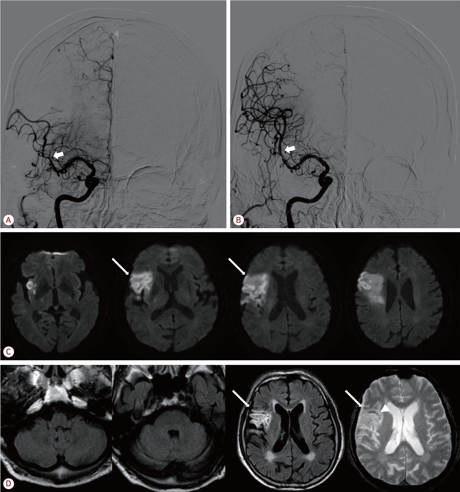
(A) Initial cerebral angiography demonstrating occlusion of the right superior M2 segment (arrow). (B) Post-procedural angiography confirming successful recanalization following mechanical thrombectomy (arrow). (C) Diffusion-weighted imaging obtained on day 2 demonstrating acute infarction predominantly involving the right insular cortex and right frontal operculum (arrows, right frontal operculum). (D) Follow-up brain MRI obtained on day 23. Fluid-attenuated inversion recovery (FLAIR) images at the levels of the medulla and pons show no new infarction in the pons or medulla, and a FLAIR image at the level of the basal ganglia shows the pre-existing right opercular- insular lesion (arrows) without new ischemic change; the corresponding gradient-echo image demonstrates mild hemorrhagic transformation within the lesion (arrowhead). MRI; magnetic resonance imaging.
By the following day, his neurological status had improved substantially, with only residual mild dysarthria and left facial palsy remaining, and his NIHSS score decreased to 2. Brain magnetic resonance imaging (MRI) performed on day 2 revealed acute infarction predominantly involving the right insular cortex and right frontal operculum (Fig. C). Electrocardiography performed during admission revealed newly detected atrial fibrillation, and anticoagulation with apixaban was subsequently initiated.
The patient had pre-existing mild exertional dyspnea attributable to his underlying HCMP. On day 6, however, he developed worsening dyspnea on exertion accompanied by audible stridor. Cardiology and pulmonology consultations, including contrast-enhanced chest CT, revealed no evidence of HCMP exacerbation, although mild pneumonia and underlying emphysema were noted; importantly, there was no mediastinal or aortic lesion and no abnormality compressing the recurrent laryngeal nerve. Serial chest radiographs likewise demonstrated a normal hilar and mediastinal contour. Serial vital sign monitoring throughout this period demonstrated no significant elevations in blood pressure or heart rate, with all values remaining within normal limits. SpO2 was maintained at 92-100% with supplemental oxygen via nasal prong, escalating from 1 to 2 L/min, and the respiratory rate ranged from 18 to 30 breaths/min. By day 9, hoarseness had become prominent alongside progressive dyspnea and stridor. Arterial blood gas analysis on 2 L/min oxygen showed pH 7.368, PaCO2 46.6 mmHg, PaO2 121.0 mmHg, and SaO2 98.0%, without hypoventilation or CO2 retention, indicating that the respiratory compromise resulted from upper airway obstruction rather than ventilatory failure. An ear, nose, and throat (ENT) specialist was consulted, and flexible fiberoptic laryngoscopy demonstrated bilateral vocal cord paralysis with only 10% airway patency at the glottic level. Immediate tracheostomy was recommended; however, the patient declined the procedure. Noting concurrent mild vocal cord edema on laryngoscopy, a 3-day course of dexamethasone was administered, and the patient was closely monitored in the intensive care unit.
Follow-up fiberoptic laryngoscopy on day 16 revealed partial improvement, demonstrating right vocal cord paralysis with left vocal cord hypomobility and airway patency recovering to 40% at the glottic level. The patient was subsequently transferred to a general ward for continued rehabilitation.
On day 23, stridor and dyspnea on exertion worsened. Repeat brain MRI showed only a mild hemorrhagic transformation within the right frontal lesion, with no evidence of new or expanded infarction, and in particular no new ischemic lesion involving the pons or medulla (Fig. D). Repeat fiberoptic laryngoscopy demonstrated deterioration of vocal cord mobility, with airway patency reduced to 20% at the glottic level. A second 3-day course of dexamethasone was administered without clinical benefit. After further counseling, the patient consented to tracheostomy, which was performed without complication. At 1-month follow-up, no meaningful recovery of vocal cord function was observed, and the patient was transferred to a rehabilitation facility with tracheostomy in place. At 1-year follow-up with ENT, the patient continued to maintain tracheostomy dependence with no significant improvement in vocal cord function.
DISCUSSION
The vocal cords are innervated by the recurrent laryngeal nerve, a branch of the vagus nerve, whose motor nucleus, the nucleus ambiguus, is located within the medulla and is known to receive bilateral corticobulbar input from both cerebral hemispheres. Consequently, a unilateral upper motor neuron lesion, including cortical infarction, is not generally expected to cause vocal cord paralysis. Nevertheless, we present an unusual case of right MCA infarction involving the opercular-insular region that subsequently led to bilateral vocal cord paralysis requiring tracheostomy.
A prior prospective study enrolling 54 acute ischemic stroke patients demonstrated that cortical infarction could produce contralateral vocal cord paresis, questioning the prevailing assumption that the nucleus ambiguus is invariably and symmetrically innervated by both cerebral hemispheres [3]. Only a few cases of bilateral vocal cord paralysis following acute ischemic stroke have been reported in the literature and the lesions in these cases commonly involved the opercular-insular region [5-7]. While the insular cortex has been implicated as a key lesion site, the adjacent frontal operculum, which harbors the laryngeal motor cortex, may act in concert with the insular cortex in mediating bilateral laryngeal motor control [4,8]. Indeed, the infarction in our patient involved the opercular-insular region as a whole, suggesting that injury to this region may be critical in disrupting bilateral vocal cord function. Taken together with these prior reports, our case raises the possibility that, in certain individuals, one hemisphere might exert dominant bilateral control over the nucleus ambiguus, such that a unilateral lesion confined to this region might be sufficient to disrupt vocal cord function on both sides.
Bilateral vocal cord paralysis in our patient developed in a delayed fashion, with symptoms emerging on day 6 and progressing through day 9, rather than at stroke onset. Reversible mechanisms such as evolving perilesional edema, progressive diaschisis, or failure of contralateral compensatory pathways may account for this delayed onset and the observed fluctuations in severity. However, these mechanisms alone do not fully explain the absence of meaningful recovery at 1-year follow-up, suggesting that permanent structural disruption of the opercular-insular corticobulbar pathways may have been the principal determinant of longterm outcome. Balagopal et al. [5] proposed autonomic dysregulation via opercular-insular cortex involvement as a potential contributor to vocal cord dysfunction in the setting of insular stroke [5,9]. However, serial vital sign monitoring in our patient demonstrated no significant elevations in blood pressure or heart rate throughout the relevant clinical period, providing no clinical evidence of sympathetic hyperactivation.
Before attributing the paralysis to the cortical infarction, more common peripheral and iatrogenic causes were considered unlikely: thrombectomy was performed under local anesthesia without airway instrumentation, the patient had no preceding neck or thoracic surgery, malignancy, or trauma, and chest imaging showed no mediastinal or aortic lesion to suggest cardiovocal (Ortner) syndrome. The delayed, fluctuating course and the absence of any new brainstem lesion on follow-up MRI further supported a supratentorial, opercular-insular mechanism. These features distinguish our case from previously reported opercular-insular cases, which were largely acute and lacked comparable exclusion of peripheral causes and follow-up imaging.
Several limitations remain. As laryngeal electromyography was not performed, a coincident peripheral lesion of the vagus or recurrent laryngeal nerve-including an age-related microvascular neuropathy-could not be entirely excluded, and the precise mechanism, including a possible autonomic contribution, remains uncertain. These limitations reflect the retrospective nature of a single case and warrant systematic electrophysiologic and autonomic evaluation in future studies.
This case underscores that bilateral vocal cord paralysis, though rarely anticipated after MCA infarction, can emerge as a delayed and life-threatening complication even following early neurological improvement. Awareness of this possibility is critical, as the subacute onset of stridor or dysphonia in the setting of opercular-insular infarction warrants prompt laryngoscopic evaluation to prevent fatal airway compromise.