중증근무력증에서 가슴샘의 역할과 병태생리

Thymic Involvement in the Pathophysiology of Myasthenia Gravis

Article information

J Korean Neurol Assoc. 2026;44(4):259-265
Publication date (electronic) : November 1, 2026
doi : http://dx.doi.org/10.17340/jkna.2026.0039
aDepartment of Neurology, Yonsei University College of Medicine, Seoul, Korea
bDepartment of Neurology, Yongin Severance Hospital, Yonsei University Health System, Yongin, Korea
정혜윤a,b, 김승우a, 신하영,a
a연세대학교 의과대학 신경과학교실
b연세대학교 의과대학 용인세브란스병원 신경과
Address for correspondence Ha Young Shin, MD, PhD Department of Neurology, Yonsei University College of Medicine, 50-1 Yonsei-ro, Seodaemun-gu, Seoul 03722, Korea Tel: +82-2-2228-1600 Fax: +82-2-393-0705 E-mail: hayshin@yuhs.ac
received : June 1, 2026 , rev-recd : June 29, 2026 , accepted : June 29, 2026 .

Trans Abstract

The precise mechanisms underlying autoantibody generation in myasthenia gravis (MG) remain incompletely understood. Evidence suggests an important role of the thymus in the initiation and maintenance of the autoimmune response. Thymic abnormalities, including thymic follicular hyperplasia and thymoma, are frequently observed in MG and are associated with distinct pathological features. In thymic hyperplasia, ectopic germinal center formation promotes B-cell activation and sustained autoantibody production, whereas thymoma is associated with impaired T-cell development and may also exhibit ectopic germinal center formation. These contribute to the dysfunction of central immune tolerance and the dysregulation of peripheral immune responses. This review summarizes the immunological mechanisms linking thymic pathology to MG pathogenesis.

서 론

중증근무력증은 자가면역질환으로 주로 신경근육접합부를 표적하는 자가항체에 의해 유발된다. 이 중 아세틸콜린수용체(acetylcholine receptor, AChR)에 대한 항체가 가장 흔하며 전체 중증근무력증 환자들의 약 85%에서 발견된다[1]. 이러한 자가항체가 형성되는 면역학적 과정은 아직 완전히 규명되지 않았으나 가슴샘이 항체 형성 과정에서 중요한 역할을 하는 것으로 알려져 있다[2].

가슴샘은 일차 림프기관으로 T림프구의 발달(development)과 선택(selection)을 담당한다[3]. 중증근무력증에서 중요한 가슴샘 병리는 두 가지로 보는데, 하나는 가슴샘증식(thymic hyperplasia)이고 다른 하나는 가슴샘종(thymoma)이다[4]. 가슴샘증식은 주로 림프구 침윤 및 구조적 과다 형성을 특징으로 하며, 특히 조기 발병 중증근무력증 환자에서 높은 빈도로 나타난다[5]. 반면 가슴샘종은 가슴샘 상피세포에서 기원하는 종양으로 중증근무력증 환자의 약 10-15%에서 가슴샘종이 발견되며[4] 가슴샘종 환자의 약 30-50%에서 중증근무력증이 동반된다[6,7]. 후자의 경우를 가슴샘종 관련 중증근무력증이라 하는데, 이는 가슴샘종이 동반되지 않은 중증근무력증에 비해 중증도가 높고 가슴샘절제술 및 종양학적 치료가 함께 고려되어야 하며 면역 억제 치료의 전략에서도 차이를 보인다[8].

치료적 관점에서도 가슴샘절제술은 중증근무력증의 주요 선택지 중 하나이다[9]. 가슴샘증식은 중증근무력증의 면역학적 기전에 관여하는 것으로 알려져 있으며 가슴샘 절제를 통해 중증근무력증 증세가 호전될 수 있다는 근거가 제시되었다[10,11]. 가슴샘종은 종양 자체의 치료를 위해 수술적 절제가 필수적이며[12], 중증근무력증의 동반 여부는 가슴종의 조직학적 아형, 수술 후 추가적인 치료 결정 등에 영향을 미친다[13].

그럼에도 불구하고 중증근무력증의 병태생리에 대해서는 여전히 해결되지 못한 영역이 많다. 자가항체 형성의 기원, 가슴샘 병터와 질환 표현형 간의 연계, 치료 반응의 이질성 등은 완전히 설명되지 않은 부분이다. 가슴샘의 면역학적 기전과 중증근무력증 발생 간의 연관성을 알아내는 것은 질환에 대한 정확한 이해를 확장하고 향후 중증근무력증의 발병 기전 규명과 치료법 개발 등의 연구 방향을 제시하는 데 중요한 의미를 가진다. 본 논문에서는 가슴샘의 면역학적 기전을 중심으로 중증근무력증 병태생리와의 연관성에 대해 종합적으로 검토하고자 한다.

본 론

1. 가슴샘의 정상 기능

가슴샘은 일차 림프기관으로 T림프구의 발생, 분화, 선택을 통해 체내 T세포 레퍼토리와 면역관용(immune tolerance)을 형성하는 데 핵심적인 역할을 한다. 골수에서 유래한 전구 T세포가 r가슴샘으로 이동하여 분화가 시작되며, 가슴샘은 피질과 수질로 구성된 영역에서 각각 서로 다른 환경을 조성하여 T세포 발달을 조절한다[14,15].

가슴샘 피질에서는 양성 선택(positive selection)이 일어난다. 이는 T세포 수용체가 주조직적합복합체(major histocompatibility complex, MHC)와 상호작용하는지 여부를 확인하는 과정이다. MHC와 적절히 결합하는 T세포만 생존하며 적절한 결합을 하지 못하는 세포는 제거된다[14].

반면 가슴샘 수질에서는 음성 선택(negative selection)과 조절 T세포(regulatory T cell, Treg) 유도가 이루어진다[15,16]. 음성 선택은 자가항원에 높은 친화도로 반응하는 자가반응(autoreactive) T세포를 제거하는 과정으로, 이 기전을 통해 중추(central) 면역관용이 형성된다. 이때 가슴샘 수질상피세포(medullary thymic epithelial cells, mTECs)는 자가면역 조절인자(autoimmune regulator, AIRE)를 통해 다양한 조직 특이 항원을 발현한다. 이렇게 T세포에게 자가항원 레퍼토리를 제시하여 자가반응 T세포의 제거를 유도한다. 중추 면역관용을 통과한 T세포는 최종적으로 CD4+ 또는 CD8+ T세포로 분화하며 말초 림프조직으로 이동하여 체내에서 면역 반응을 담당하게 된다. 가슴샘 수질에서는 특정 자가항원에 대한 비교적 높은 친화도의 T세포 수용체 신호를 받은 일부 T세포가 음성 선택으로 제거되는 대신 Treg으로의 분화 과정을 거칠 수 있으며, 이런 과정은 말초 면역 조절 유지에 중요한 역할을 한다.

2. 중증근무력증의 면역 병태생리

중증근무력증의 가장 흔한 병인 항체는 AChR에 대한 항체이며, 이외에도 근육특이인산화효소(muscle specific kinase, MuSK) 및 저밀도지단백질관련단백질4 (low-density lipoprotein receptor-related protein 4, LRP4)에 대한 항체도 보고된다[1].

AChR항체는 여러 기전을 통해 신경근육접합부의 장애를 유발한다[17]. 첫째는 주로 immunoglobulin G1 (IgG1)과 immunoglobulin G3 (IgG3) 아형으로 알려져 있는 AChR항체가 보체 의존 세포독성을 유도하여 막공격복합체를 형성하고 신경근육접합부의 구조적 파괴를 초래하는 기전이다. 둘째로, 항체가 AChR의 리간드결합부위에 결합하여 아세틸콜린과 수용체의 결합을 직접적으로 방해하는 기능적 차단 기전이 있다. 셋째로, AChR항체에 의한 수용체 교차결합이 이루어져 AChR의 내재화와 분해가 유도된다[18-20].

반면 MuSK에 대한 항체는 병태생리 기전이 AChR항체와 차이점이 있다. MuSK 단백질은 신경근육접합부에서 agrin-LRP4-MuSK 복합체를 형성하여 AChR의 군집화와 신경근육접합부의 구조적 안정성을 유지하는 데 필수적인 역할을 한다[21]. MuSK에 대한 자가항체는 복합체의 형성을 교란하여 AChR 군집화와 신호전달을 저해한다. 특히 MuSK항체는 immunoglobulin G4 (IgG4) 아형으로 AChR항체와 달리 보체 활성화나 항체 매개 세포독성을 유발하기보다는 단백질 간의 상호작용을 차단하는 기전이 중심이 되는 것으로 알려져 있다[22]. 한편 LRP4항체에 의해 유발되는 중증근무력증도 보고되었으나 정확한 병태생리는 아직 잘 알려져 있지 않다[23].

중증근무력증에서 자가항체 생성은 B세포의 비정상 분화와 활성화 과정으로 이는 자가항원 특이 CD4+ T세포와의 상호 작용에 의해 매개된다[24]. 특히 소포-도움T세포(follicular helper T cell, Tfh)와의 반응을 통해 B세포는 종류전환재조합(classswitch recombination)과 체세포 과돌연변이를 거쳐 병인성 자가항체를 생성하는 형질세포로 분화한다[2,24-27]. 이렇게 생성된 형질세포는 항체 역가를 지속적으로 유지하여 자가면역질환의 만성화를 설명하는 중요한 기전이다.

한편 중증근무력증에서는 자가항체 생성을 적절히 제어하는 면역 억제 기전의 결함이 동반된다. T도움17 (T helper 17, Th17)세포의 증가와 Treg의 감소 또는 기능 저하가 관찰되었으며[24], 이런 과정은 B세포 활성과 항체 생성 반응을 간접적으로 증폭시키는 요인이 된다. Th17세포가 염증사이토카인을 분비하여 면역 활성화와 조직 내 염증 반응을 촉진하는 반면 자가반응림프구를 억제하여 말초 면역관용을 유지해야 하는 Treg의 가능은 약화되어 있다. 결과적으로 Th17세포와 Treg의 불균형은 중증근무력증에서 B세포 매개 자가항체 반응이 효과적으로 제어되지 못하는 환경을 제공하며 질환의 지속성과 중증도를 결정하는 중요한 병태생리 요소로 작용한다[2,24].

3. 가슴샘증식과 중증근무력증

가슴샘소포증식(thymic follicular hyperplasia)은 중증근무력증에서 가장 흔하게 관찰되는 가슴샘 이상으로, 특히 조기 발병 중증근무력증 환자에서 높은 빈도로 나타난다[5]. 조직학적으로는 림프구 침윤의 증가, 피질과 수질의 구조 재편성, 종자 중심(germinal center, GC)의 형성을 특징으로 한다(Fig. 1) [28]. 이러한 변화는 가슴샘이 T세포 성숙 기관의 기능을 넘어 자가면역 반응이 지속적으로 증폭되는 비정상 면역 활성화 공간으로 기능함을 시사한다.
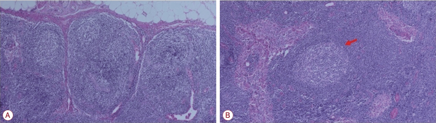
Figure 1.

(A) Histopathological features of thymic follicular hyperplasia (H&E, ×40) showing compartmentalized thymic architecture with lymphoid follicle formation. (B) Higher magnification demonstrating a well-developed germinal center (red arrow; H&E, ×80). H&E; hematoxylin and eosin.

가슴샘증식에서 가장 특징적인 병리학적 소견은 GC의 형성이다. 정상 가슴샘에서는 GC가 거의 관찰되지 않으나 중증근무력증 환자의 가슴샘증식 조직에서는 림프절과 유사한 구조를 갖는 GC가 다수 형성된다[5]. GC는 항원 특이 B세포가 Tfh세포와의 상호작용을 통하여 증식, 분화, 항체 친화도 성숙이 이루어지는 미세환경이다. GC 내에서는 특히 가슴샘 내 근육모양세포(myoid cell)에서 발현되는 AChR을 포함한 자가항원이 제시되며, 이에 특이적으로 반응하는 B세포가 형질세포로 분화한다. 이 과정에서 종류전환재조합(classswitch recombination)과 체세포 과돌연변이를 통해 병인성 자가형체가 생성된다[29-31]. GC 형성과 유지는 Tfh세포와 밀접하게 연관된다. 가슴샘증식 조직에서는 Tfh세포의 증가와 interleukin 21 (IL-21), CXCL13 등 사이토카인과 케모카인의 발현이 증가하며 이는 B세포의 GC 유입에 기여한다[31,32]. 이를 종합하였을 때 흉선비대 내 GC는 자가항체 생성의 중요한 구조로 작용한다.

또한 가슴샘증식은 AIRE 기능 이상과도 연관된다. AIRE는 mTECs에서 조직특이항원의 발현을 유도하여 자가반응 T세포의 음성 선택을 매개하는 핵심 인자이다[33,34]. 일부 연구에 따르면 가슴샘증식 조직 내 AIRE 발현 감소, 조직특이항원 제시 이상, 항원제시세포 기능 변화 등이 관찰되며 이러한 변화는 정상 가슴샘에서 이루어져야 할 중추 면역관용의 저하로 이어질 수 있다[34]. AIRE는 자가항원 특이 Treg의 선택과 형성에 필수적인 역할을 수행하므로 AIRE의 발현 감소는 Treg의 생성과 안정성을 저해하여 가슴샘 내 중추 면역관용의 붕괴에 기여한다[35,36]. 가슴샘증식 조직에서 Treg의 감소와 면역 억제 기능 저하가 보고되었으며, 이는 자가반응 림프구의 조절 실패로 이어져 지속적인 자가면역 활성화를 야기한다[37]. 결과적으로 자가반응 T세포의 탈출과 항원 특이 Treg세포의 감소를 통해 면역 조절 시스템의 불균형을 유발하며, 이는 CD4+ T세포의 비정상 분화를 촉진하게 된다. 따라서 가슴샘증식 조직 내에서는 자가항원 특이 CD4+ T세포가 지속적으로 활성화되어 있는데 IL-6, IL-21, IL-23, transforming growth factor-β (TGF-β)와 같은 사이토카인 신호는 CD4+ T세포가 Th17 세포로 분화되도록 유도한다. Th17세포는 IL-17과 같은 염증 사이토카인을 분비하여 국소 염증 반응과 항원제시기능을 증폭시키고 B세포 활성화를 강화한다[38-40].

이런 면역학적 변화는 중증근무력증의 임상적 특징과도 밀접하게 연관된다. 가슴샘증식은 주로 AChR항체 양성인 중증근무력증 환자들에서 관찰되며 비교적 젊은 연령에서 발병하고 항체의 역가가 높은 경향을 보인다. 이런 환자군에서는 면역 억제 치료와 함께 빠른 가슴샘절제술이 치료 전략으로 활용된다[5,10,11]. 가슴샘절제술은 병리적 GC 제거와 비정상 항원 제시 환경의 교정을 통해 자가항체 생성을 감소시키며, 이러한 면역학적 변화는 질병 증상의 호전, 약물 요구량 감소 및 관해율 증가와 연관되는 것으로 보고되었다.

4. 가슴샘종과 중증근무력증

가슴샘종(thymoma)은 가슴샘의 상피세포에서 기원하는 종양으로 중증근무력증 환자의 약 10-15%에서 발견된다[4,6]. 가슴샘종 관련 중증근무력증은 앞서 언급한 가슴샘증식에서의 중증근무력증과는 일부 다른 병태생리 기전을 가지며 중증도가 높은 임상 양상을 보이는 것이 특징이다[41,42]. 가슴샘종의 내부 미세환경은 정상적인 T세포 분화를 수행하는 데 구조적, 기능적 결함이 있는 것으로 알려져 있다. 우선 가슴샘종 내의 종양 상피세포는 정상적인 피질과 수질 구조를 형성하지 못하여 T세포가 성숙 과정에서 거쳐야 하는 해부학적 구획화가 무너진다[6]. 특히 가슴종 조직에서는 AIRE의 발현이 낮거나 결핍되어 있으며 MHC class II 분자의 발현 또한 저하되어 있다[43,44]. 이는 조직특이항원의 제시와 음성 선택이 제대로 작동하지 않음을 의미하며 중추 면역관용의 붕괴로 자가반응 T세포들이 미성숙한 상태로 말초 혈액으로 방출되는 병리적 상황을 야기한다.

가슴샘종 내의 이런 상황은 자가항체의 스펙트럼에서도 차이를 만든다. 가슴샘종 환자들에서는 제1형 인터페론(interferon, INF; INF-α, IFN-ω)이나 IL-12와 같은 사이토카인에 대한 자가항체가 발견된다. 이는 가슴샘종 내 복잡한 면역 환경을 시사한다.45 또한 가슴샘종 관련 중증근무력증에서는 titin, ryanodine receptor와 같은 가로무늬근육 항체가 높은 빈도로 확인된다[13,46].

가슴샘종에서 T세포에 대한 면역 교육의 부전은 세계보건기구(World Health Organization, WHO) 조직학적 분류에 따른 아형별 특징과 연관이 있다. Type B2 가슴샘종은 중증근무력증 동반율이 가장 높은 것으로 보고되어 있는 반면 type A나 C의 경우 중증근무력증 발생 빈도가 상대적으로 낮다[47]. 이는 가슴샘 피질에서 다량의 미성숙 T세포를 유인하고 보유하는 특징을 보이는 type B2 가슴샘종에서 자가반응 T세포가 더 많이 생성될 가능성이 있기 때문으로 생각된다. 반면 상피세포의 비정형성이 두드러지고 T세포 유입이 적은 type A나 type C에서는 가슴샘 내 T세포와 상피세포 사이의 상호작용이 제한적이기 때문에 자가반응 T세포가 형성될 가능성이 상대적으로 낮아 중증근무력증의 발생 빈도가 낮은 것으로 추정된다[48,49].

또 하나의 주목할 부분은 종양 자체뿐만 아니라 가슴샘종 주변부의 조직학적 변화이다. 가슴샘종을 둘러싼 비종양성 가슴샘 조직에서는 가슴샘증식과 유사하게 GC의 형성이 관찰되기도 한다(Fig. 2) [50,51]. 이런 주변부의 GC는 종양 내부에서 교육에 실패하고 방출된 자가반응 T세포가 B세포를 자극하여 자가항체 생성을 증폭시키는 과정에 기여할 가능성이 있다. 즉 가슴샘종 관련 중증근무력증은 종양 내부의 중추 면역관용 붕괴와 주변부 조직의 비정상 GC 형성이 결합된 복합적인 병태생리 결과물이라고 볼 수 있다.
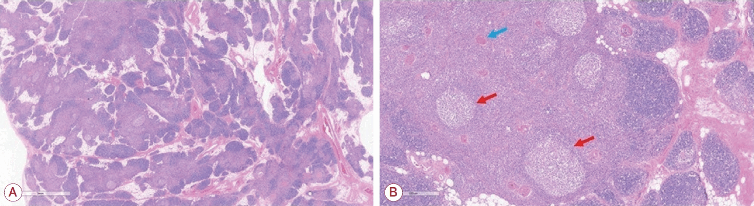
Figure 2.

(A) Representative hematoxylin and eosin (H&E)-stained sections of non-neoplastic tissue from a patient with type B2 thymoma (H&E, ×20). Preserved lobulated thymic architecture separated by fibrous septa with numerous lymphoid follicles. (B) Higher magnification of representative germinal centers from panel A demonstrates well-formed germinal centers (red arrows; H&E, ×40). Hassall corpuscles (blue arrow) are present, demonstrating preserved non-neoplastic thymic tissue.

이러한 병리적 구조는 가슴샘종 절제 시 종양뿐만 아니라 주변의 잔여 가슴샘 조직까지 광범위하게 제거해야 하는 임상적 근거가 된다[41]. 가슴샘종은 종양 자체의 치료를 위해 수술적 절제가 필수적이지만 수술 후에도 중증근무력증 증상이 지속되거나 오히려 악화되는 경우 또는 새롭게 중증근무력증이 발생하는 경우가 보고된다[52-55]. 이는 종양 내에 존재하던 자가반응 T세포들이 종양을 제거하면서 일시적으로 말초로 유출되거나 수술 시 완전히 제거되지 않은 주변부의 활성화된 면역 조직들이 자가항체 생성을 지속하는 것이 원인이 될 수 있다. 또한 수술 전 이미 말초로 유출되어 골수 등에 위치하는 장수 형질세포의 기여로도 설명될 수 있다. 이들은 가슴샘이 제거된 후에도 장기간 자가항체를 생성하여 보체 활성화와 신경근육접합부의 손상 및 AChR 감소를 유도하고 지속시킬 수 있다. 따라서 가슴샘절제술 이후에도 일정 기간 동안 중증근무력증 증상이 지속되거나 일시적으로 악화될 수 있으며 수술의 완전한 효과가 나타나기까지 시간적 지연이 발생할 수 있다[56,57]. 최근 연구에서 가슴샘종 주변 조직에서 발견되는 GC가 수술 후 발생하는 중증근무력증과 연관될 수 있음을 보고하였으며, 이러한 가슴샘종 주변부 면역 환경이 질환 발생에 기여할 가능성을 제시하였다[58].

결 론

중증근무력증의 병태생리에서 가슴샘은 단순히 자가항체가 생성되는 장소를 넘어 중추 면역관용의 붕괴와 자가반응 림프구의 증폭이 일어나는 중요한 면역학적 거점이다. 조기 발병 중 증근무력증에서 주로 관찰되는 가슴샘증식은 GC 형성과 B세포 활성화를 촉진하는 염증 미세환경을 통해 자가면역 반응을 지속시킨다. 이와 달리 가슴샘종 관련 중증근무력증은 종양 상피세포의 종양적 변화로 가슴샘 내 미세환경 변화와 AIRE 결핍으로 인한 음성 선택의 실패가 특징이다. 또한 종양 내부의 T세포 교육 부전뿐만 아니라 가슴샘종 주변부 조직에서 비정상 GC 형성을 동반하여 자가항체 생성의 복합적인 병태생리를 형성할 가능성이 시사된다.

최근 신생아Fc수용체억제제 및 보체억제제 등 새로운 생물학적 제제들이 도입되며 중증근무력증의 치료 틀이 급격하게 변화하고 있다[9,59]. 하지만 이 표적 치료제들은 중증근무력증을 조절하는 데에는 효과적이지만 중증근무력증을 완치하지는 못한다. 중증근무력증에서 자가면역 반응의 초기 과정이 이루어지는 장소인 가슴샘의 병태생리학 기전을 정확히 규명할 수 있다면 중증근무력증의 면역학 근간을 보다 정확하게 이해하고 향후 중증근무력증에 대한 맞춤형 치료 설계 및 질병의 완치를 이루는 토대가 될 것으로 기대된다.

References

1. Gilhus NE, Tzartos S, Evoli A, Palace J, Burns TM, Verschuuren J. Myasthenia gravis. Nat Rev Dis Primers 2019;5:30.
2. Berrih-Aknin S, Le Panse R. Myasthenia gravis: a comprehensive review of immune dysregulation and etiological mechanisms. J Autoimmun 2014;52:90–100.
3. Thapa P, Farber DL. The role of the thymus in the immune response. Thorac Surg Clin 2019;29:123–131.
4. Gilhus NE, Verschuuren JJ. Myasthenia gravis: subgroup classification and therapeutic strategies. Lancet Neurol 2015;14:1023–1036.
5. Truffault F, de Montpreville V, Eymard B, Sharshar T, Le Panse R, Berrih-Aknin S. Thymic germinal centers and corticosteroids in myasthenia gravis: an immunopathological study in 1035 cases and a critical review. Clin Rev Allergy Immunol 2017;52:108–124.
6. Scorsetti M, Leo F, Trama A, D’Angelillo R, Serpico D, Macerelli M, et al. Thymoma and thymic carcinomas. Crit Rev Oncol Hematol 2016;99:332–350.
7. Romi F, Hong Y, Gilhus NE. Pathophysiology and immunological profile of myasthenia gravis and its subgroups. Curr Opin Immunol 2017;49:9–13.
8. Romi F, Gilhus NE, Aarli JA. Myasthenia gravis: clinical, immunological, and therapeutic advances. Acta Neurol Scand 2005;111:134–141.
9. Narayanaswami P, Sanders DB, Wolfe G, Benatar M, Cea G, Evoli A, et al. International consensus guidance for management of myasthenia gravis: 2020 update. Neurology 2021;96:114–122.
10. Wolfe GI, Kaminski HJ, Aban IB, Minisman G, Kuo HC, Marx A, et al. Randomized trial of thymectomy in myasthenia gravis. N Engl J Med 2016;375:511–522.
11. Wolfe GI, Kaminski HJ, Aban IB, Minisman G, Kuo HC, Marx A, et al. Long-term effect of thymectomy plus prednisone versus prednisone alone in patients with non-thymomatous myasthenia gravis: 2-year extension of the MGTX randomised trial. Lancet Neurol 2019;18:259–268.
12. Falkson CB, Bezjak A, Darling G, Gregg R, Malthaner R, Maziak DE, et al. The management of thymoma: a systematic review and practice guideline. J Thorac Oncol 2009;4:911–919.
13. Romi F. Thymoma in myasthenia gravis: from diagnosis to treatment. Autoimmune Dis 2011;2011:474512.
14. Marx A, Yamada Y, Simon-Keller K, Schalke B, Willcox N, Strobel P, et al. Thymus and autoimmunity. Semin Immunopathol 2021;43:45–64.
15. Cheng M, Anderson MS. Thymic tolerance as a key brake on autoimmunity. Nat Immunol 2018;19:659–664.
16. Owen DL, Sjaastad LE, Farrar MA. Regulatory T cell development in the thymus. J Immunol 2019;203:2031–2041.
17. Dresser L, Wlodarski R, Rezania K, Soliven B. Myasthenia gravis: epidemiology, pathophysiology and clinical manifestations. J Clin Med 2021;10:2235.
18. Drachman DB, Angus CW, Adams RN, Michelson JD, Hoffman GJ. Myasthenic antibodies cross-link acetylcholine receptors to accelerate degradation. N Engl J Med 1978;298:1116–1122.
19. Hara H, Hayashi K, Ohta K, Itoh N, Nishitani H, Ohta M. Detection and characterization of blocking-type anti-acetylcholine receptor antibodies in sera from patients with myasthenia gravis. Clin Chem 1993;39:2053–2057.
20. Rodgaard A, Nielsen FC, Djurup R, Somnier F, Gammeltoft S. Acetylcholine receptor antibody in myasthenia gravis: predominance of IgG subclasses 1 and 3. Clin Exp Immunol 1987;67:82–88.
21. Kim N, Stiegler AL, Cameron TO, Hallock PT, Gomez AM, Huang JH, et al. Lrp4 is a receptor for agrin and forms a complex with MuSK. Cell 2008;135:334–342.
22. Huijbers MG, Zhang W, Klooster R, Niks EH, Friese MB, Straasheijm KR, et al. MuSK IgG4 autoantibodies cause myasthenia gravis by inhibiting binding between MuSK and Lrp4. Proc Natl Acad Sci U S A 2013;110:20783–20788.
23. Plomp JJ, Huijbers MG, van der Maarel SM, Verschuuren JJ. Pathogenic IgG4 subclass autoantibodies in MuSK myasthenia gravis. Ann N Y Acad Sci 2012;1275:114–122.
24. Uzawa A, Kuwabara S, Suzuki S, Imai T, Murai H, Ozawa Y, et al. Roles of cytokines and T cells in the pathogenesis of myasthenia gravis. Clin Exp Immunol 2021;203:366–374.
25. Klein U, Dalla-Favera R. Germinal centres: role in B-cell physiology and malignancy. Nat Rev Immunol 2008;8:22–33.
26. Vinuesa CG, Sanz I, Cook MC. Dysregulation of germinal centres in autoimmune disease. Nat Rev Immunol 2009;9:845–857.
27. Allen CD, Okada T, Cyster JG. Germinal-center organization and cellular dynamics. Immunity 2007;27:190–202.
28. Levinson AI, Wheatley LM. The thymus and the pathogenesis of myasthenia gravis. Clin Immunol Immunopathol 1996;78:1–5.
29. Sims GP, Shiono H, Willcox N, Stott DI. Somatic hypermutation and selection of B cells in thymic germinal centers responding to acetylcholine receptor in myasthenia gravis. J Immunol 2001;167:1935–1944.
30. Zuckerman NS, Howard WA, Bismuth J, Gibson K, Edelman H, Berrih-Aknin S, et al. Ectopic GC in the thymus of myasthenia gravis patients show characteristics of normal GC. Eur J Immunol 2010;40:1150–1161.
31. Sikorski PM, Kaminski HJ, Kusner LL. Thymic hyperplasia in myasthenia gravis: a narrative review. Mediastinum 2025;9:17.
32. Anderson MS, Venanzi ES, Chen Z, Berzins SP, Benoist C, Mathis D. The cellular mechanism of AIRE control of T cell tolerance. Immunity 2005;23:227–239.
33. Gardner JM, Fletcher AL, Anderson MS, Turley SJ. AIRE in the thymus and beyond. Curr Opin Immunol 2009;21:582–589.
34. Passos GA, Speck-Hernandez CA, Assis AF, Mendes-da-Cruz DA. Update on AIRE and thymic negative selection. Immunology 2018;153:10–20.
35. Malchow S, Leventhal DS, Nishi S, Fischer BI, Shen L, Paner GP, et al. Aire-dependent thymic development of tumor-associated regulatory T cells. Science 2013;339:1219–1224.
36. Perry JSA, Lio CJ, Kau AL, Nutsch K, Yang Z, Gordon JI, et al. Distinct contributions of AIRE and antigen-presenting-cell subsets to the generation of self-tolerance in the thymus. Immunity 2014;41:414–426.
37. Fattorossi A, Battaglia A, Buzzonetti A, Ciaraffa F, Scambia G, Evoli A. Circulating and thymic CD4 CD25 T regulatory cells in myasthenia gravis: effect of immunosuppressive treatment. Immunology 2005;116:134–141.
38. Villegas JA, Bayer AC, Ider K, Bismuth J, Truffault F, Roussin R, et al. IL-23/Th17 cell pathway: a promising target to alleviate thymic inflammation maintenance in myasthenia gravis. J Autoimmun 2019;98:59–73.
39. Dong C. Differentiation and function of pro-inflammatory Th17 cells. Microbes Infect 2009;11:584–588.
40. Waite JC, Skokos D. Th17 response and inflammatory autoimmune diseases. Int J Inflam 2012;2012:819467.
41. Masaoka A, Yamakawa Y, Niwa H, Fukai I, Kondo S, Kobayashi M, et al. Extended thymectomy for myasthenia gravis patients: a 20-year review. Ann Thorac Surg 1996;62:853–859.
42. Budde JM, Morris CD, Gal AA, Mansour KA, Miller JI Jr. Predictors of outcome in thymectomy for myasthenia gravis. Ann Thorac Surg 2001;72:197–202.
43. Strobel P, Chuang WY, Chuvpilo S, Zettl A, Katzenberger T, Kalbacher H, et al. Common cellular and diverse genetic basis of thymoma-associated myasthenia gravis: role of MHC class II and AIRE genes and genetic polymorphisms. Ann N Y Acad Sci 2008;1132:143–156.
44. Marx A, Hohenberger P, Hoffmann H, Pfannschmidt J, Schnabel P, Hofmann HS, et al. The autoimmune regulator AIRE in thymoma biology: autoimmunity and beyond. J Thorac Oncol 2010;5 Suppl 4:S266–S272.
45. Meager A, Wadhwa M, Dilger P, Bird C, Thorpe R, Newsom-Davis J, et al. Anti-cytokine autoantibodies in autoimmunity: preponderance of neutralizing autoantibodies against interferon-alpha, interferon-omega and interleukin-12 in patients with thymoma and/or myasthenia gravis. Clin Exp Immunol 2003;132:128–136.
46. Romi F, Bo L, Skeie GO, Myking A, Aarli JA, Gilhus NE. Titin and ryanodine receptor epitopes are expressed in cortical thymoma along with costimulatory molecules. J Neuroimmunol 2002;128:82–89.
47. Kumar R. Myasthenia gravis and thymic neoplasms: a brief review. World J Clin Cases 2015;3:980–983.
48. Okumura M, Fujii Y, Shiono H, Inoue M, Minami M, Utsumi T, et al. Immunological function of thymoma and pathogenesis of paraneoplastic myasthenia gravis. Gen Thorac Cardiovasc Surg 2008;56:143–150.
49. Marx A, Willcox N, Leite MI, Chuang WY, Schalke B, Nix W, et al. Thymoma and paraneoplastic myasthenia gravis. Autoimmunity 2010;43:413–427.
50. Lefeuvre CM, Payet CA, Fayet OM, Maillard S, Truffault F, Bondet V, et al. Risk factors associated with myasthenia gravis in thymoma patients: the potential role of thymic germinal centers. J Autoimmun 2020;106:102337.
51. Berrih-Aknin S, Morel E, Raimond F, Safar D, Gaud C, Binet JP, et al. The role of the thymus in myasthenia gravis: immunohistological and immunological studies in 115 cases. Ann N Y Acad Sci 1987;505:50–70.
52. Yamada Y, Yoshida S, Iwata T, Suzuki H, Tagawa T, Mizobuchi T, et al. Risk factors for developing postthymectomy myasthenia gravis in thymoma patients. Ann Thorac Surg 2015;99:1013–1019.
53. Sun XG, Wang YL, Liu YH, Zhang N, Yin XL, Zhang WJ. Myasthenia gravis appearing after thymectomy. J Clin Neurosci 2011;18:57–60.
54. Kondo K, Monden Y. Myasthenia gravis appearing after thymectomy for thymoma. Eur J Cardiothorac Surg 2005;28:22–25.
55. Kim A, Choi SJ, Kang CH, Lee S, Son H, Kim JA, et al. Risk factors for developing post-thymectomy myasthenia gravis in patients with thymoma. Muscle Nerve 2021;63:531–537.
56. Jiang R, Hoehn KB, Lee CS, Pham MC, Homer RJ, Detterbeck FC, et al. Thymus-derived B cell clones persist in the circulation after thymectomy in myasthenia gravis. Proc Natl Acad Sci U S A 2020;117:30649–30660.
57. Berrih-Aknin S, Ragheb S, Le Panse R, Lisak RP. Ectopic germinal centers, BAFF and anti-B-cell therapy in myasthenia gravis. Autoimmun Rev 2013;12:885–893.
58. Chung HY, Shin HY, Choi YC, Park HJ, Lee JG, Lee CY, et al. Germinal centers are associated with postthymectomy myasthenia gravis in patients with thymoma. Eur J Neurol 2024;31:e16119.
59. Gilhus NE, Andersen H, Andersen LK, Boldingh M, Laakso S, Leopoldsdottir MO, et al. Generalized myasthenia gravis with acetylcholine receptor antibodies: a guidance for treatment. Eur J Neurol 2024;31:e16229.

Article information Continued

Figure 1.

(A) Histopathological features of thymic follicular hyperplasia (H&E, ×40) showing compartmentalized thymic architecture with lymphoid follicle formation. (B) Higher magnification demonstrating a well-developed germinal center (red arrow; H&E, ×80). H&E; hematoxylin and eosin.

Figure 2.

(A) Representative hematoxylin and eosin (H&E)-stained sections of non-neoplastic tissue from a patient with type B2 thymoma (H&E, ×20). Preserved lobulated thymic architecture separated by fibrous septa with numerous lymphoid follicles. (B) Higher magnification of representative germinal centers from panel A demonstrates well-formed germinal centers (red arrows; H&E, ×40). Hassall corpuscles (blue arrow) are present, demonstrating preserved non-neoplastic thymic tissue.