섬망을 보인 환자에서 관찰된 광범위하게 확장된 혈관주위공간: 스위스 치즈 양상

Widespread Enlarged Perivascular Spaces in a Patient with Delirium: Swiss Cheese Appearance

Article information

J Korean Neurol Assoc. 2024;42(4):433-434
Publication date (electronic) : November 1, 2024
doi : http://dx.doi.org/10.17340/jkna.2024.0023
aDepartment of Neurology, National Cancer Center, Goyang, Korea
bDepartment of Neurology, Inje University Sanggye Paik Hospital, Seoul, Korea
김기훈,a,b, 정승호b, 김수현a, 현재원a, 김호진a
a국립암센터 신경과
b인제대학교상계백병원 신경과
Address for correspondence Ki Hoon Kim, MD Department of Neurology, Inje University Sanggye Paik Hospital, 1342 Dongil-ro, Nowon-gu, Seoul 01757, Korea Tel: +82-2-950-1090 Fax: +82-2-950-1908 E-mail: p6p6gogo@naver.com
received : March 11, 2024 , rev-recd : July 3, 2024 , accepted : July 4, 2024 .

유방암으로 항암 치료를 진행하던 59세 여성에서 섬망이 발생하였다. 뇌자기공명영상 검사에서 양측 대뇌 전반에 조영증강이 되지 않는 다발성 낭종이 관찰되었는데 액체감쇠역전회복영상(fluid attenuated inversion recovery)에서 뇌척수액과 등음영(isodense)을 보이고 자화율강조영상(susceptibility weighted imaging)에서는 저신호강도는 없었다(Fig.). 뇌척수액 검사는 정상이었고 바이러스, 세균, 결핵균, 곰팡이, 기생충을 포함한 균 검사와 세포(cytology) 검사는 음성이었다. 섬망 호전 후에 시행한 간이정신상태 검사(Korean version of mini-mental state examination, K-MMSE)는 25점, 임상치매평가척도(clinical dementia rating scale, CDR)는 0.5점이었다. 신경심리 검사에서 집중력, 기억력, 시공간능력, 전두엽 기능의 저하를 보였다. 확장된 혈관주위공간(enlarged perivascular spaces)은 대뇌소혈관질환(cerebral small vessel disease)과의 연관성이 보고되었고 고령 혹은 신경퇴행질환에서 흔하게 관찰된다[1]. 반면 본 증례에서 보인 광범위하게 확장된 혈관주위공간(widespread enlarged perivascular spaces)은 굉장히 드물게 발생하고 특징적인 영상 소견 때문에 “Swiss cheese brain”으로 기술되기도 하였다[2]. 대부분은 무증상이나 일부 환자에서 인지장애나 국소적인 신경계 이상이 동반되었다[3,4]. 아직 발병 기전과 임상적인 의의는 잘 알려져 있지 않고 증례에서 관찰된 인지 저하와의 관련성도 현 시점에서는 불명확하다.
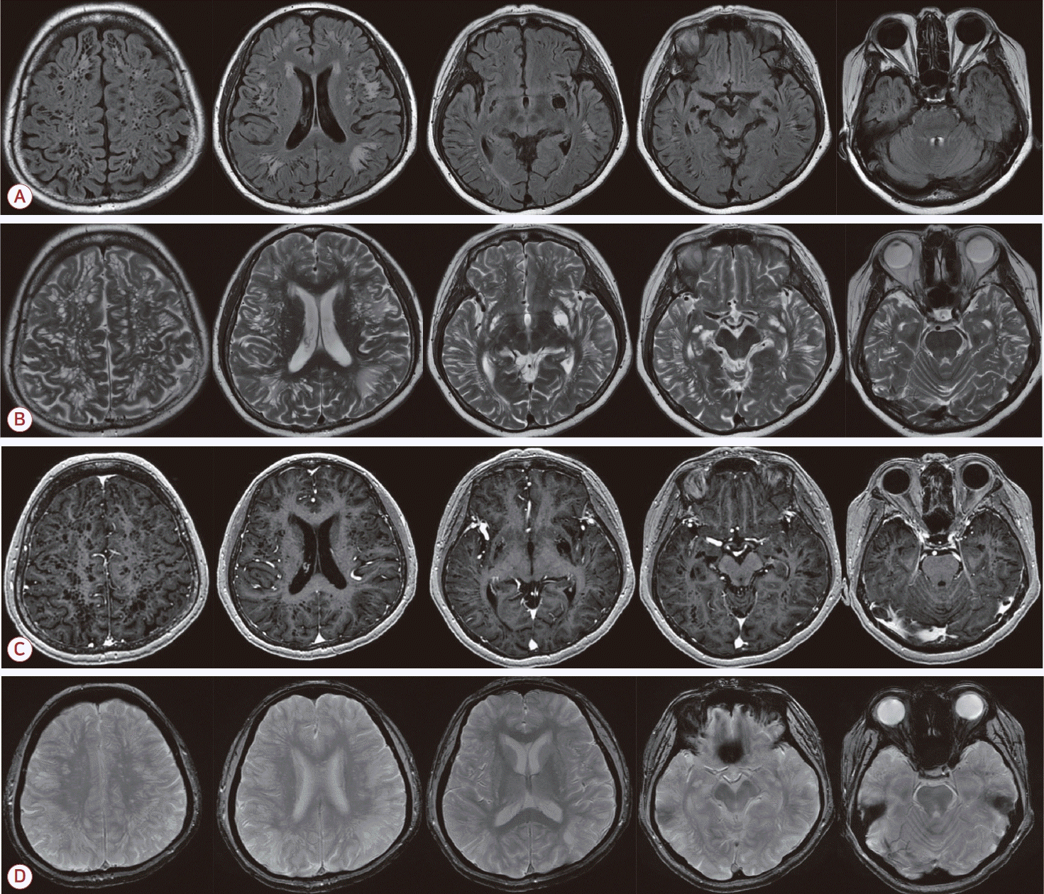
Figure.

Brain MRI showed widespread multiple and cystic lesions in the bilateral cerebral hemisphere on FLAIR and T2-weighted imaging (A, B), and gadolinium enhancement imaging (C). These cystic lesions were isointense to CSF, encircled by a rim of hyperintensities with no gadolinium enhancement. SWI showed no sign of focal hemorrhage and hemosiderosis (D). MRI; magnetic resonance imaging, FLAIR; fluid- attenuated inversion recovery, CSF; cerebrospinal fluid, SWI; susceptibility weighted image.

References

1. Moon WJ. Preface for imaging of neurodegenerative diseases: current status and recent updates. J Korean Soc Radiol 2022;83:451–452.
2. Cochrane TI, Nguyen TN. Swiss cheese brain. Neurology 2007;68:140.
3. Maclullich AM, Wardlaw JM, Ferguson KJ, Starr JM, Seckl JR, Deary IJ. Enlarged perivascular spaces are associated with cognitive function in healthy elderly men. J Neurol Neurosurg Psychiatry 2004;75:1519–1523.
4. Taniguchi D, Shimura H, Watanabe M, Hattori N, Urabe T. Widespread enlarged perivascular spaces associated with dementia and focal brain dysfunction: case report. BMC Neurol 2017;17:210.

Article information Continued

Figure.

Brain MRI showed widespread multiple and cystic lesions in the bilateral cerebral hemisphere on FLAIR and T2-weighted imaging (A, B), and gadolinium enhancement imaging (C). These cystic lesions were isointense to CSF, encircled by a rim of hyperintensities with no gadolinium enhancement. SWI showed no sign of focal hemorrhage and hemosiderosis (D). MRI; magnetic resonance imaging, FLAIR; fluid- attenuated inversion recovery, CSF; cerebrospinal fluid, SWI; susceptibility weighted image.