J Korean Neurol Assoc > Volume 42(4); 2024 > Article
일과성경동맥주위염증증후군: 혈관벽영상 및 조영증강초음파를 통한 진단적 통찰을 포함한 증례 보고

Abstract

Transient perivascular inflammation of the carotid artery (TIPIC) syndrome is a rare disease accompanied by acute neck pain and specific pathologic changes in the carotid artery and surrounding tissue. Here, we present a case diagnosed with typical imaging features, including extensive enhancement with luminal narrowing on vessel wall imaging and the presence of microbubbles on contrast-enhanced ultrasonography. These findings provide valuable insights into the pathophysiology of TIPIC syndrome and importance of imaging modalities for early detection and management.

일과성경동맥주위염증(transient perivascular inflammation of the carotid artery, TIPIC)증후군은 경동맥 분기부 주위에 갑작스러운 통증이 발생하는 드문 질환이다. 이전 보고에서 급성 경부통증 환자 중 약 2% 정도의 유병률을 보고하였으며[1] 국내 보고는 매우 드물다. 발생 원인과 병태생리가 알려져 있지 않기 때문에 초기에 진찰과 병력 청취로 진단하기 어렵다. 급성 경부통증 환자에서 TIPIC증후군을 진단하기 위해서는 경동맥박리, 갑상샘염, 자가면역 또는 감염 등에 의한 혈관염, 침샘염(sialadenitis), 경추질환 등을 감별해야 한다. TIPIC증후군은 다른 원인 없이 특징적인 편측 급성 경부 통증과 경동맥 주위의 염증 반응이 확인되고 자가 회복하는 경과를 보일 경우 진단할 수 있다. 본 증례는 다중영상방식(multimodality imaging)을 통해 확인한 TIPIC증후군의 특징적 신경영상에 대해 보고한다.

증 례

51세 남자가 3일 전 시작된 좌측 경부의 묵직한 통증으로 병원에 왔다. 최근 외상, 발열 등은 없었고 박동성 통증이 지속되었다. 좌측 경동맥이 위치한 부위의 동통, 발적, 부종이 확인되었다. 혈액 검사에서 백혈구, 적혈구침강속도(erythrocyte sedimentation rate) 및 프로칼시토닌(procalcitonin)은 정상 범위였으며, C-반응단백질(C-reactive protein)이 1.92 mg/L (정상, 0.00-0.22)로 증가되어 있었다.
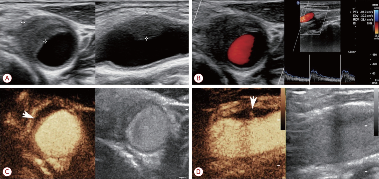
초음파 검사 B-mode에서 좌 측 경동맥 분지에 표층으로 치우친 균질한 저음영 병변(eccentric homogeneous hypoechoic lesion)과 40% 협착이 확인되었다(Fig. 1-A). 혈관내막(tunica intima)과 중간막(tunica media)은 구분이 뚜렷하지 않고 Doppler mode에서 최대 수축기 속도(peak systolic velocity)는 정상이었다(Fig. 1-B). 조영제(SonoVueTM; Bracco, Milano, Italy) 2 mL를 주입 후 시행한 조영증강초음파(contrast-enhanced ultrasonography) 검사에서는 중간막의 국소적 조영증강과 혈관 중간-바깥막의 조영증강되는 미세기포가 관찰되었다(Fig. 1-C, D).
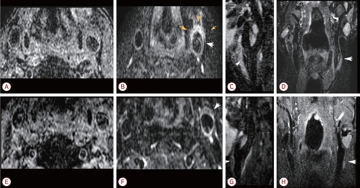
자기공명혈관벽영상(high resolution magnetic resonance vessel wall imaging, HR-VWI)의 조영증강 전 T1강조영상에서 병변 부위는 전체적으로 저신호강도를 보였다(Fig. 2-A). 조영증강 후 혈관외막(tunica adventitia)과 내막은 뚜렷하게 T1 조영증강되었으며 중간막은 증강되지 않고 팽창되어 있었다. 또한 혈관주위조직 부종 부위는 조영증강되었다(Fig. 2-B-D).
환자는 2주간의 비스테로이드소염제와 스테로이드 치료를 병행하였고 증상 발생 7일 후부터 부종과 통증은 완화되었다. 3개월 추적 관찰에서 뇌졸중 등 허혈성 질환 없이 완전히 회복되었고 영상 검사에서 경미한 혈관벽의 비후가 관찰되었다(Fig. 2-E-H).

고 찰

일측성 경부통증은 1988년에 Fay에 의해 carotidynia로 명명되었지만[2] 그 모호함과 광범위함으로 말미암아 2004년 International Headache Society의 두통 분류에서 제외되었다. 그러나 유사한 사례들이 지속적으로 보고되고 영상 검사의 발달로 경동맥과 주위 조직의 침범이 실재하는 것으로 확인되면서 TIPIC증후군으로 그 범례가 굳어졌다[1]. TIPIC증후군은 본 증례처럼 급성 통증을 동반하는 원인 불명의 비감염 염증부종이 경동맥과 주위 조직에 발생하여 혈관벽 비후와 일시적인 혈관 협착이 생기는 것이 영상 검사로 확인되며 감염이나 종괴, 자가면역질환 등이 배제되면 진단을 내릴 수 있다[1]. 대부분 양성 경과를 보이기 때문에 병리 소견에 대해서는 알려진 바가 적었으며 국내에서는 질환의 확인도 거의 없었다.
TIPIC증후군의 양성 경과를 고려할 때 본 증례와 같은 영상 검사가 초기 진단에 도움이 될 수 있다. 다만 영상 검사에서 경동맥의 협착, 염증과 주변 조직의 광범위한 침윤이 확인되기 때문에 루프스, 자가면역갑상샘염(autoimmune thyroiditis) 등의 자가면역질환, 경동맥박리, 혈관염, 외상, 국소 감염에 대한 감별이 필요하다[3-5].
TIPIC증후군은 신경영상에서 경동맥 내부 및 주변 조직의 침범이 확인된다. 경동맥 혈관 중간막과 외막의 침범이 흔하며[6,7] MR-VWI가 해상도가 높아 내막과 중간막을 구분하는 데 도움이 된다. 경동맥 주변의 지방조직이 연조직으로 대체되며 관찰되는 혈관주변침윤(perivascular infiltration)은 computed tomography, magnetic resonance imaging (MRI), 초음파 모두에서 관찰 가능하다. 염증 반응의 활동성 및 범위에 대해서는 조영증강 MRI 또는 초음파 검사가 도움이 될 수 있다[4,5].
TIPIC증후군은 경부에 발생하는 매우 드문 질환이지만 임상적 예후는 대부분 양호하다. 그럼에도 불구하고 대뇌의 혈류를 공급하는 경동맥 부위에 발생하는 급성, 미만성 염증 반응인 점을 고려할 때 신속하고 정확한 검사를 통해 위험한 질환에 대해 감별이 반드시 필요하다. 본 증례에서는 TIPIC증후군의 전형적인 임상 경과를 보이는 환자에서 관찰되는 다양한 신경영상 검사 결과를 확인할 수 있었다. TIPIC증후군에서 이러한 영상 검사는 초기 진단 및 경과의 추적 관찰에 도움이 되는 유용한 검사이다.

REFERENCES

1. Lecler A, Obadia M, Savatovsky J, Picard H, Charbonneau F, de Champfleur NM, et al. TIPIC syndrome: beyond the myth of carotidynia, a new distinct unclassified entity. AJNR Am J Neuroradiol 2017;38:1391-1398.
crossref pmid pmc
2. Fay T. Atypical neuralgia. Arch Neurol Psychiatry 1927;18:309-315.

3. Rafailidis V, Chryssogonidis I, Tegos T, Partovi S, Charitanti-Kouridou A, Staub D. Role of multi-parametric ultrasound in transient perivascular inflammation of the carotid artery syndrome. Ultrasound 2019;27:77-84.
crossref pmid pmc
4. Micieli E, Voci D, Mumoli N, Mastroiacovo D, Grigorean A, Obadia M, et al. Transient perivascular inflammation of the carotid artery (TIPIC) syndrome. Vasa 2022;51:71-77.
crossref pmid
5. Scoppettuolo P, Dalaqua M, Ghika JA, Bonvin C. Multimodal imaging for a TIPIC syndrome case. Acta Neurol Belg 2022;122:1069-1071.
crossref pmid
6. Kim HS, Woo JS, Kim BY, Jang HH, Hwang SJ, Kwon SJ, et al. Biochemical and clinical correlation of intraplaque neovascularization using contrast-enhanced ultrasound of the carotid artery. Atherosclerosis 2014;233:579-583.
crossref pmid
7. Ulus S, Ozcan UA, Arslan A, Buturak A, Dincer A, Kara S, et al. Imaging spectrum of TIPIC syndrome: validation of a new entity with vessel wall imaging. Clin Neuroradiol 2020;30:145-157.
crossref pmid

Figure 1.
Ultrasonography shows luminal narrowing and intralesional microbubble after enhancement. (A) The transverse and longitudinal view of ultrasonography images show eccentric perivascular infiltration as hypoechoic mural thickening at the level of the carotid artery bifurcation, and a moderate lumen narrowing. (B) There was no hemodynamic change in Doppler mode. After a bolus injection of 2.5 mL SonoVueTM (Bracco, Milano, Italy), transverse (C) and longitudinal view (D) of CEUS images (left, contrast mode; right, two-dimensional mode) show enhanced microbubbles within the lesion (arrowhead), suggesting non-specific inflammatory process. CEUS; contrast-enhanced ultrasound.
jkna-42-4-357f1.jpg
Figure 2.
Comparison of initial (A-D) and 3 months post-onset MR-VWI (E-H). (A) Initial MP-RAGE image shows anterolateral luminal narrowing of left common carotid artery with low signal intensity. (B-D) Contrast-enhanced T1 weighted image shows low signal intensity core lesion and extensively enhanced soft plaque (white arrowhead). Perivascular enhancement is also noted (yellow arrowheads). (E) MP-RAGE and (F-H) contrast-enhanced T1 weighted image performed 3 months after symptom onset show resolved luminal narrowing, decreased extent of perivascular infiltration and size of intimal flap (white arrowhead). MR-VWI; magnetic resonance vessel wall imaging, MP-RAGE; magnetization prepared rapid gradient echo.
jkna-42-4-357f2.jpg
TOOLS
METRICS Graph View
  • 0 Crossref
  •  0 Scopus
  • 111 View
  • 14 Download
Related articles


ABOUT
BROWSE ARTICLES
EDITORIAL POLICY
FOR CONTRIBUTORS
Editorial Office
(ZIP 03163) #1111, Daeil Bldg, 12, Insadong-gil, Jongno-gu, Seoul, Korea
Tel: +82-2-737-6530    Fax: +82-2-737-6531    E-mail: jkna@neuro.or.kr                

Copyright © 2024 by Korean Neurological Association.

Developed in M2PI

Close layer