동정맥샛길에 의한 안면마비
Infratemporal Arteriovenous Fistula Presenting with Peripheral Facial Palsy
Article information
Trans Abstract
Peripheral facial palsy results from facial nerve injury. While most cases are idiopathic (Bell's palsy) with favorable prognosis, other causes such as trauma, infection, and compression have poor outcomes. An arteriovenous fistula (AVF) is a direct connection between an artery and a vein that is treatable with endovascular procedures, making early diagnosis essential. We report a case of a 68-year-old woman with peripheral facial palsy and pulsatile pain for four months, diagnosed with an infratemporal AVF compressing the facial nerve.
말초안면마비는 한쪽 얼굴근육마비를 주요 증상으로 하며 안면신경 손상으로 인해 발생한다[1]. 대부분의 경우 특발성(벨마비)이지만 청신경종이나 신경초종과 같은 종양, 중이염이나 꼭지돌기염과 같은 염증, 외상이나 바이러스 감염, 동맥류나 동정맥샛길과 같은 혈관 이상에 의한 압박으로도 발생할 수 있다[2]. 벨마비는 6개월 이내에 대부분 회복되어 예후가 양호한 반면 압박 원인으로 인한 경우 예후가 좋지 않다[3]. 동정맥샛길은 선천 또는 후천 원인에 의해 동맥과 정맥 사이에 모세혈관을 우회하는 미세한 연결이 생기는 혈관 이상이며 혈관 내 시술로 치료가 가능한 질병으로 정확한 조기 진단이 중요하다[4]. 본 논문에서는 동정맥샛길로 인해 말초안면마비가 동반된 증례를 보고하고자 한다.
증 례
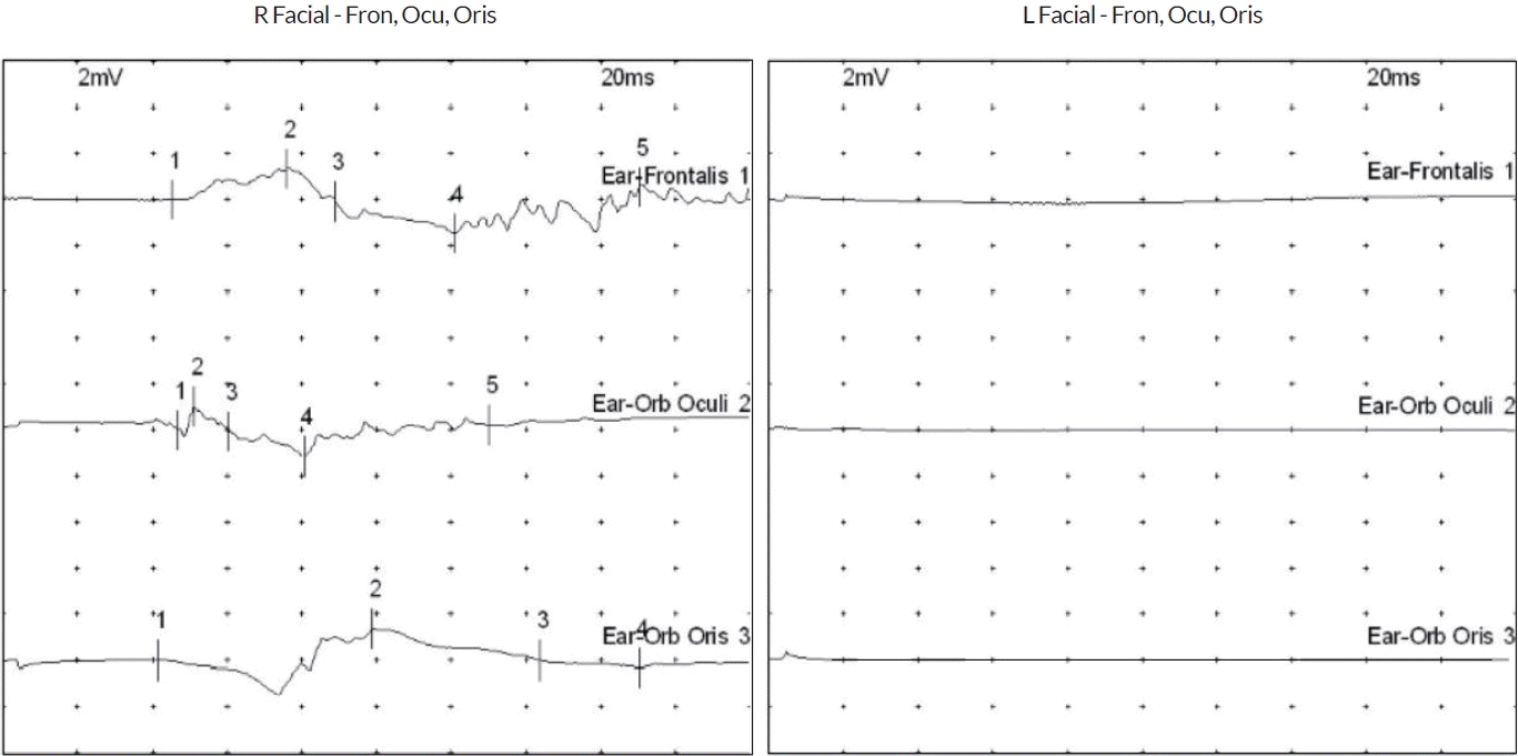
68세 여자가 4개월 전부터 갑자기 발생한 말초안면마비와 안면 통증으로 병원에 왔다. 혈관 위험 인자를 포함한 과거 병력은 없었다. 좌측 턱 밑부터 후두부로 이어지는 박동통증과 해당 부위 열감과 부종을 동반하였다. 좌측 이마 주름이 지어지지 않았고 좌측 눈꺼풀과 입꼬리가 처지는 말초안면마비를 보였다(Fig. 1-A). 환자는 다른 병원에서 벨마비로 진단되어 메틸프레드니솔론 8 mg 하루 2회 주사 치료를 5일 받았다. 일반 혈액 검사, 혈청전해질 검사 그리고 혈액 화학 검사에서 이상은 없었으며 수두대상포진바이러스 immunoglobulin (Ig) G 양성, IgM 음성이었다. 안면신경전도 검사에서 좌측 이마근, 눈둘레근, 입둘레근에서 복합근육활동전위가 확인되지 않았다(Fig. 2). 스테로이드 치료 후에도 안면마비의 호전을 보이지 않았으며 좌측 턱 밑과 후두부의 통증이 지속되어 별신경절차단을 반복 시행하였으나 호전이 없었다.

(A) Facial image. (B) Time-of-flight (TOF) image shows enhancement on infratemporal area. (C) Magnetic resonance angiography (MRA) showing engorged veins on arterial phase, left infratemporal area and internal jugular vein (IJV). (D) left anteroposterior and (E) lateral transfemoral cerebral angiography showing arteriovenous fistula fed from left external carotid artery (ECA), drained to IJV (orange arrow). (F) Cerebral angiography showing distal internal maxillary artery, middle deep temporal artery, middle meningeal artery, and superficial temporal artery with a reduced flow of arteriovenous fistula after embolization (orange arrow).

Facial NCS were conducted on 25 days after symptom onset. NCS showed no compound muscle action potential on the left frontalis, orbicularis oculi/oris muscles. Fron; frontalis, Ocu; orbicularis oculi, Oris; orbicularis oris, NCS; nerve conduction study.
추가적인 평가를 위해 신경과에 방문하였고 안면신경 압박 병터 여부를 확인하기 위해 뇌자기공명영상 및 자기공명혈관 조영영상을 시행하였으며 측두하 부위에 동정맥샛길이 확인되었다(Fig. 1-B, C). 혈관조영술을 시행하였고 좌측 원위부 내 상악동맥, 중간심부측두동맥, 중간수막동맥, 표재측두동맥에서 상악동을 지나 내경정맥까지 이어진 동정맥샛길을 확인하였다. 정맥역류는 관찰되지 않았다(Fig. 1-D, E) 동정맥샛길의 크기와 분포를 고려하였을 때 좌측 안면신경의 무릎신경절부터 말단가지가 분지하기 전까지의 부위를 압박하였을 것이라고 판단하였다. 이후 코일색전술을 시행하였고(Fig. 1-F) 2주 정도 경과한 뒤부터 안면마비 및 통증은 호전 양상을 보였으며 현재 별다른 합병증 없이 경과 관찰 중에 있다.
고 찰
본 증례는 중년 여성에서 갑자기 발생한 좌측 턱 밑부터 후두부로 이어지는 박동통증과 동반된 좌측 말초안면마비로 병원에 와서 스테로이드 치료 및 물리 치료를 받았으나 호전을 보이지 않은 경우이다. 압박 병터를 의심하여 영상 검사를 시행하였고 안면신경을 압박하는 좌측 외경동맥에서 내경정맥으로 빠져나가는 동정맥샛길을 확인할 수 있었다.
안면신경은 다리뇌뒤판에서 시작하여 소뇌다리뇌각을 지나 측두골 내부로 진입한 후 내이도를 통과하여 안면신경관의 무릎신경절을 지나 붓꼭지구멍을 통해 두개 외 연부조직 구역으로 진입한다. 이후 두개 외 연부조직에서 붓돌기의 전외측으로 주행하며 귀밑샘을 통과한 후 각각의 종말부로 분지된다[5]. 보고에 의하면 말초안면신경마비 중 75%는 벨마비로 진단되고 감염, 종양, 자가면역질환이 원인이 되는 경우가 종종 있으나 혈관 이상에 의한 압박이 원인인 경우는 흔치 않다[6]. 일반적인 벨마비의 경우 스테로이드 치료와 항바이러스 치료를 병행하고 70-90%에서는 3-7일 이내에 회복을 보이기 시작하나 기타 다른 원인으로 인하여 발병할 경우 약물 치료에 반응을 보이지 않기 때문에 2주 이상 증상이 지속되는 말초안면마비의 경우 다른 원인 감별에 대한 고려가 필요하다[7].
또한 환자가 호소하는 통증의 양상과 정도는 벨마비 이외 말초안면마비의 다른 원인을 감별하는 데 도움이 된다[8]. 일반적으로 벨마비는 주로 귓바퀴 뒤 부위에 경미하거나 중등도의 통증을 동반하며 심한 경우 찌릿한 느낌의 신경통 양상으로 나타난다[8]. 그러나 본 증례의 환자는 안면 부위의 열감 및 부종을 동반한 강한 박동통증을 호소하였고 스테로이드 치료에도 반응이 없었다. 과거 연구에 의하면 동정맥샛길은 생성되는 위치에 따라 신경을 압박하여 이명이나 안신경, 안면신경마비 등을 일으킬 수 있고 67%의 환자에서 박동두통을 호소하는데 동정맥샛길의 위치에 따라 통증 호소 부위가 다르다[9]. 통증 부위를 고려할 때 본 환자에서 보인 해당 부위의 박동통증은 안면신경이 주행하는 측두하 부위의 동정맥샛길로 인한 압박 병터에서 기인한다고 볼 수 있다.
과거 외상 이후 발생한 중수막동정맥샛길이 얼굴신경의 주행 부위에 발생하면서 안면신경 압박에 의해 말초안면마비가 발생하였던 증례가 있었으며 수술 처치를 시행하고 5일 후부터 신경계 증상의 호전을 보였다[10]. 하지만 본 증례의 환자는 특별한 외상이나 유발 요인 없이 좌측 측두하 부위의 동정맥샛길이 확인되었고 증상 발생부터 진단까지 오랜 시간이 걸렸으며 시술 후부터 증상의 호전이 앞선 증례보다 늦었다는 점에서 환자의 임상 증상과 경과에 따른 빠른 진단이 필요하다는 점을 시사한다.
본 증례는 안면마비 외에 심한 박동통증을 동반할 경우 추가적인 평가를 통해 동정맥샛길에 의한 압박 병터를 고려해야 함을 보여준다. 동정맥샛길은 치료가 가능한 질환이기 때문에 빠른 진단이 필요하다. 아직까지 국내에서는 별다른 유발 요인 없이 발생한 동정맥샛길로 인해 말초안면마비와 통증을 호소한 환자에서 시술 후 신경계 증상의 호전을 보인 증례가 보고된 바 없어 저자들은 본 환자를 보고하고자 한다.