아급성뇌경색에서 세포독성부종을 동반한 Wallerian 변성

Wallerian Degeneration with Cytotoxic Edema in the Subacute Stage of Cerebral Infarction

Article information

J Korean Neurol Assoc. 2022;40(2):200-201
Publication date (electronic) : May 1, 2022
doi : http://dx.doi.org/10.17340/jkna.2022.2.21
Department of Neurology, Catholic University of Daegu School of Medicine, Daegu, Korea
김요셉, 도영록
대구가톨릭대학교 의과대학 신경과학교실
Address for correspondence: Youngrok Do, MD Department of Neurology, Catholic University of Daegu School of Medicine, 33 Duryugongwon-ro 17-gil, Nam-gu, Daegu 42472, Korea Tel: +82-53-650-3064 Fax: +82-53-654-9786 E-mail: dyr4173@cu.ac.kr
received : October 1, 2021 , rev-recd : December 15, 2021 , accepted : December 15, 2021 .

79세 여성이 내원 2개월 전 발생한 보행장애가 서서히 진행되었다. 내원 1개월 전부터는 안정 떨림이 동반되었다. 편마비는 없었다. 내원 시 자기공명영상에서 왼쪽 기저핵에 아급성 뇌경색이 있었다(Fig.). 왼쪽 내포와 중뇌 대뇌각에 확산강조영상 고신호, 겉보기 확산 계수지도 저신호의 세포독성부종 병변이 있었는데, 증상이 지속적으로 악화되었고 병변이 기저핵 병변으로부터 하행 피질 척수로에 위치하며 서로 다른 혈관 영역(territory)에 위치하므로 뇌경색의 재발이 아닌 뇌경색 후 진행한 아급성기의 Wallerian 변성(Wallerian degeneration)으로 생각된다.

Wallerian 변성에 관한 이전 보고들은 뇌경색에서 확산강조영상에서 고신호이면서 겉보기 확산 계수 등/고신호의 Wallerian 변성이거나[1] 뇌출혈 또는 천막 밑 경색에서의 보고였으나[2] 본 증례는 천막위 뇌경색에서 천막 밑으로 이어지는 겉보기 확산 계수 저신호의 아급성기 Wallerian 변성이었다. 본 증례를 통해 Wallerian 변성은 아급성기 뇌경색에서 세포독성부종을 동반할 수 있을 것으로 생각한다.
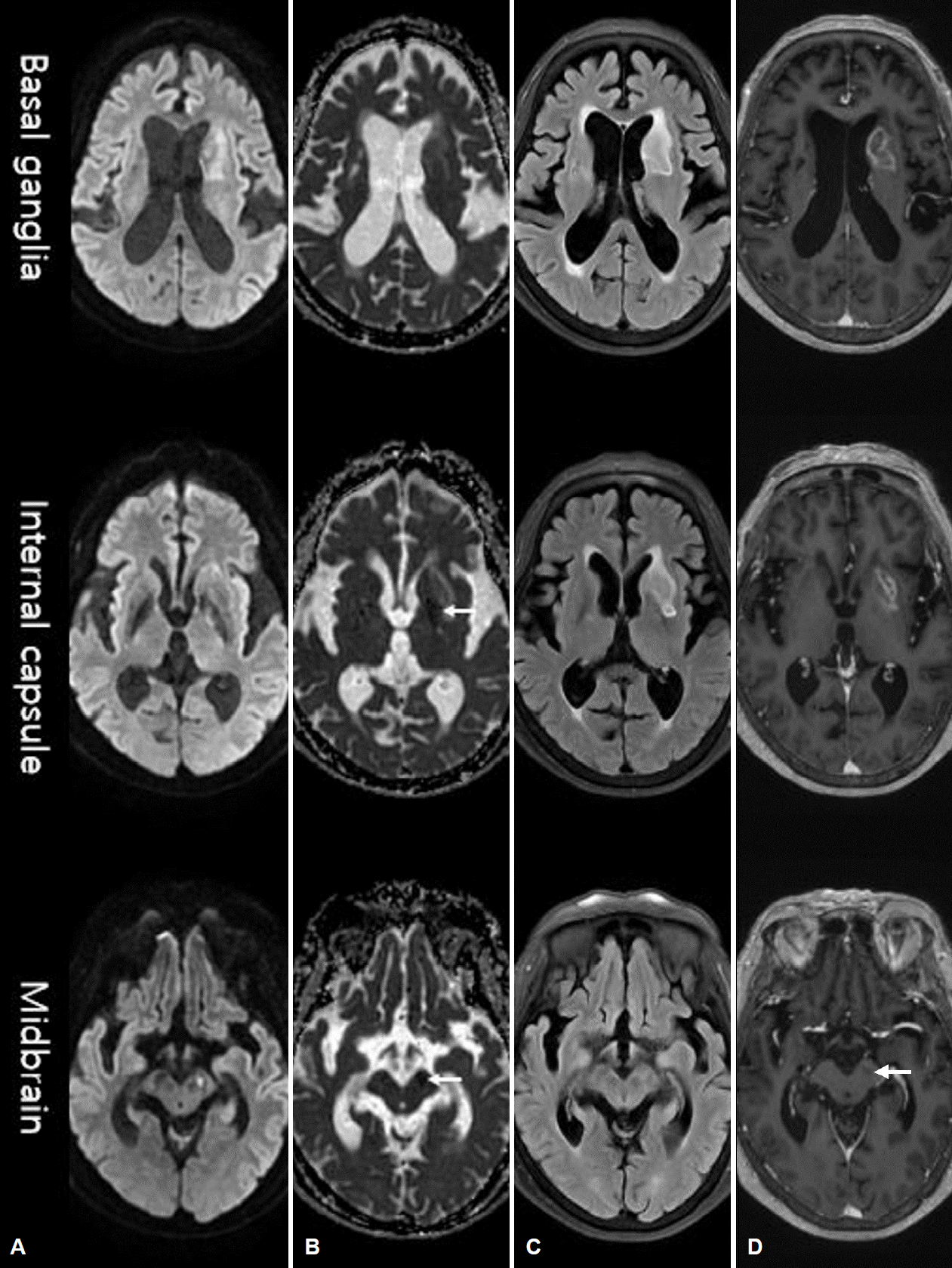
Figure.

Brain magnetic resonance imaging of the patient. Images are shown at basal ganglia, internal capsule, and midbrain level consecutively. The left basal ganglia had a subacute infarction which showed iso to high signal on apparent diffusion coefficient (ADC) images. Simultaneously, (B) cytotoxic edema lesions located long downstream along the corticospinal tract from the basal ganglia infarction showed low signals (arrows) (D) without enhancement in the left internal capsule and midbrain cerebral peduncle, in different vascular territories. (A) Diffusion-weighted image; (B) ADC; (C) fluid attenuated inversion recovery; (D) T1 enhanced.

References

1. Zheng X, Zhang Y, Man Y, Hu Z, Zhang N, Pan S. Clinical features, risk factors, and early prognosis for Wallerian degeneration in the descending pyramidal tract after acute cerebral infarction. J Stroke Cerebrovasc Dis 2021;30:105480.
2. Musson R, Romanowski C. Restricted diffusion in Wallerian degeneration of the middle cerebellar peduncles following pontine infarction. Pol J Radiol 2010;75:38–43.

Article information Continued

Figure.

Brain magnetic resonance imaging of the patient. Images are shown at basal ganglia, internal capsule, and midbrain level consecutively. The left basal ganglia had a subacute infarction which showed iso to high signal on apparent diffusion coefficient (ADC) images. Simultaneously, (B) cytotoxic edema lesions located long downstream along the corticospinal tract from the basal ganglia infarction showed low signals (arrows) (D) without enhancement in the left internal capsule and midbrain cerebral peduncle, in different vascular territories. (A) Diffusion-weighted image; (B) ADC; (C) fluid attenuated inversion recovery; (D) T1 enhanced.