쇄골하동맥으로의 잘못된 중심정맥관 삽입으로 인하여 발생한 카테터 주변 혈전증 및 뇌경색증
Inadvertent Subclavian Artery Catheterization: Juxta-Catheter Thrombosis with Subsequent Stroke
Article information
케모포트(chemoport)는 피하로 삽입되는 쇄골하 중심정맥관의 일종으로 중환자, 특히 화학요법(chemotherapy)을 받는 환자들에게 약제를 중심정맥을 통하여 안정적이며 간편하게 투입하는 방법으로 널리 사용되고 있다. 케모포트 삽입에 필요한 중심정맥 천자 과정은 단순하고 안전한 방법으로 여겨지나 기흉, 혈흉, 심장눌림증(cardiac tamponade), 혈관 손상, 혈전 또는 색전증 등의 드물지만 심각한 부작용들이 보고되어 있다[1]. 그중에서 동맥으로의 잘못된 천자(arterial catheterization)는 중심정맥관 삽입술 과정 중 1% 미만에서 발생하는 비교적 드문 합병증으로, 혈전 형성 및 이로 인한 뇌경색이 발생하는 사례가 보고된 바 있으나 아직 국내에는 보고된 적이 없다[1]. 저자들은 쇄골하동맥(subclavian artery)으로의 잘못된 천자로 인한 쇄골하동맥 내 혈전 증식 및 이로 인한 후방순환영역 뇌경색이 발생한 사례를 경험하여 보고한다.
증 례
직장암으로 진단받은 55세 여자가 수술 절제술 후 화학요법을 위하여 외부 병원에서 케모포트 삽입술을 받았다. 이후 일상생활에 불편함 없이 지내오던 중 시술 10일 후 비교적 갑자기 발생한 1시간 동안의 일시적인 우측 다리 위약감과 구음장애가 발생하여 본원 응급실로 내원하였다. 내원 후 시행한 신체검사 및 신경학적 진찰에서는 특이 소견이 없었으나 확산강조영상(diffusion-weighted image)에서 우측 내측 측두엽(medial temporal lobe), 시상(thalamus) 및 우측 소뇌에 급성 뇌경색을 시사하는 다발성 고신호 강도 병변이 확인되었다(Fig. A-C). 대동맥 상방 전산화단층혈관조영술(supra-aortic computed tomography angiography)에서 우측 쇄골하동맥으로 케모포트 도관이 잘못 삽입되어 있는 것이 확인되었고(Fig. D) 그 주변으로 혈관내 혈전으로 의심되는 이상 음영이 확인되었다(Fig. E). 색전성 뇌경색(embolic cerebral infarction)의 원인으로 동맥내 카테터 주위 혈전 형성이 우선적으로 고려되어 저분자량헤파린(low molecular weight heparin, nadroparin 2,850 IU)을 환자에게 하루 두 번 피하 주사하였다. 일주일 후 추적검사로 시행한 흉부 전산화단층촬영혈관조영술(chest computed tomography angiography, CTA)에서 도관 주변에 확인되던 혈전의 크기가 감소된 것을 확인할 수 있었다(Fig. F). 뇌경색의 원인인 케모포트 및 이와 연결된 중심정맥관을 제거하기로 결정하였으며, 제거술은 스텐트 이식편 보조(stent graft assistance) 하에 시술과 관련된 합병증 없이 성공적으로 수행되었다. 입원 기간 중 수행한 신경학적 진찰에서 국소 신경계증상은 확인되지 않았다. 환자는 퇴원 후 아스피린(100 mg/day)을 1년간 복용하였다. 이후 시행한 대동맥 상방 CTA 추적 영상에서 잔여 혈전은 확인되지 않았으며(Fig. G), 추적관찰 기간 동안 허혈뇌졸중을 시사하는 증상은 없었다.
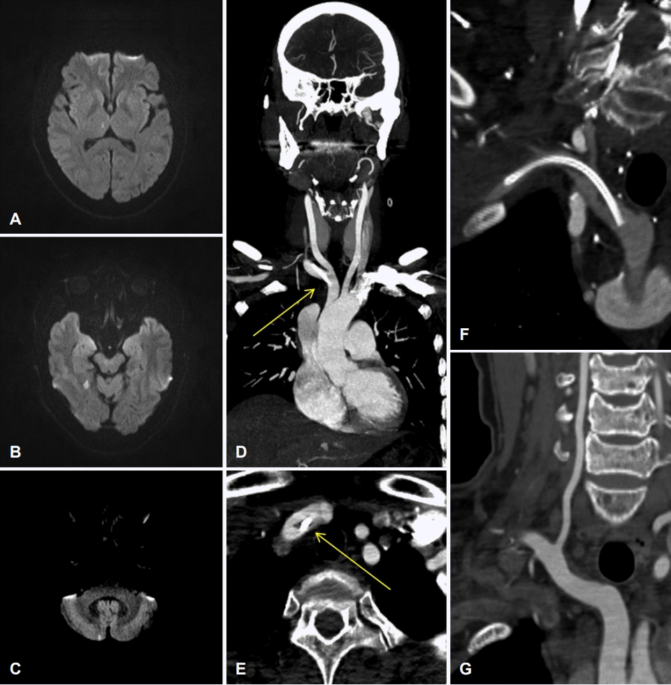
(A-C) Brain diffusion-weighted image on the day of admission showing multiple patchy hyperintensities on the right thalamus, right medial temporal lobe, and right cerebellum. (D, E) Supra-aortic CTA showing central venous catheter inadvertently placed on right subclavian artery and suspected juxta-catheter thrombosis (arrows). (F) One-week follow-up chest CTA after anticoagulation showing reduction of thrombosis around the catheter. (G) One-year follow-up supra-aortic CTA showing no visible thrombosis on right subclavian artery. CTA; computed tomography angiography.
고 찰
본 증례에서 쇄골하동맥으로 잘못 삽입된 중심정맥도관에서 형성된 혈전으로 인하여 유발된 허혈뇌졸중의 드문 사례를 소개하였다. 현재까지 보고된 바에 의하면 중심정맥관 내부 및 주변의 혈전 형성은 보고에 따라 8-63%의 발생률을 보인다[1-3]. 의료기관에 따라 차이가 있지만 중심정맥관 삽입 시 일반적으로 빗장뼈의 정중선(midline of clavicle) 등의 해부학적 표지자(anatomical landmark)에 따라 천자 위치를 정한 후, 천자 이후 혈류의 박동성 여부에 따라 동-정맥을 구분 하는 방법이 통용되고 있다[4]. 하지만 심장기능이 저하된 만성 질환을 지닌 환자에서 동-정맥 간의 박동성 차이는 간과될 수 있으며[5,6], 해부학적 표지자에 기반한 중심정맥관 삽입술 시 시술의 성공률은 94%, 동맥 천자율은 4%에 이른다는 보고가 있다[4]. 정맥 카테터의 잘못된 동맥 삽입을 방지하기 위하여 현재까지 이루어진 연구 중 초음파 유도 중심정맥관 삽입(ultrasound-guided central line insertion)이 동맥 천자의 위험을 효과적으로 줄이는 것으로 나타났다[4,7]. 한편, 동맥 천자의 결과로 발생한 혈전에 대하여 항응고요법의 지속 기간과 구체적인 약제의 종류에 대하여 정립된 치료 방법은 없으며, 본 증례와 같이 혈전으로 인한 급성 허혈뇌졸중이 동반된 경우 출혈변환(hemorrhagic conversion)의 위험으로 약제 사용 및 그 기간에 있어 논쟁의 여지가 있다. 하지만 추가적인 혈전 증식 및 이로 인한 뇌경색 발생의 예방에 있어 각 환자에게 개별화된 방법으로 혈전 형성의 원인이 된 카테터의 즉각적 제거 및 빠른 시간 내 항응고 치료는 중요할 것으로 판단된다.