임상 추론: 두통과 복시로 내원한 38세 남자
Clinical Reasoning: A 38-year-old Man Presented with Headache and Diplopia
Article information
증 례
38세 남자가 두통과 복시로 응급실에 왔다. 두통은 6일 전부터 발생하였고 좌측 눈과 좌측 이마에 국한되었으며 욱씬거리는 양상이었다. 두통 발생 3일 후부터는 수평복시가 발생하였다. 복시는 한쪽 눈씩 가릴 때는 소실되었고, 멀리 보거나 좌측으로 주시할 때 심해졌으며 일중 변동 없이 지속되었다. 고혈압이나 당뇨, 갑상선 질환의 병력은 없었으며, 최근 감염이나 발열도 없었다. 신체 진찰에서 눈꺼풀뒤당김(lid retraction)이나 안구돌출(exophthalmos) 등은 보이지 않았고 신경학적 진찰에서 좌측 눈의 외전장애가 관찰되었다. 하지만 내전, 상전, 하전은 정상이었고, 우측 눈의 운동도 정상이었다(Fig. 1). 시력, 동공반사를 포함한 다른 뇌 신경검사는 정상이었고 시신경유두부종 등의 이상 소견은 관찰되지 않았으며, 뇌수막자극징후도 없었다.
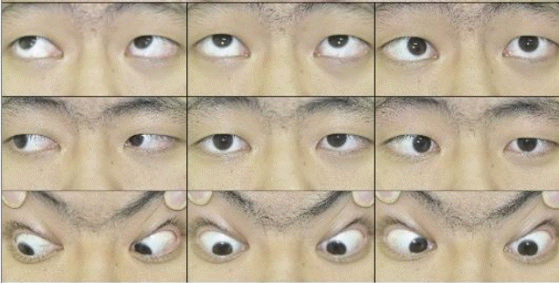
Nine gaze photographs show left esotropia in the neurtral position and abduction deficit of the left eye.
질문 1. 병변의 위치는?
신경학적 진찰에서 좌측 눈의 외전장애가 관찰되었기 때문에, 병변의 위치는 외안근이나 신경근접합부 이상 또는 외전신경(abducens nerve)을 생각해 볼 수 있다(Fig. 2). 외안근 병변으로 발생하는 질환은 갑상선눈병증이나 안근염, 종양 침윤 등이 있으며, 신경근접합부병변으로 발생하는 질환으로는 중증근무력증이 대표적이다[1]. 외전신경은 교뇌와 연수 사이를 빠져나와 Dorello관을 통하여 해면정맥동(cavernous sinus)으로 들어가며, 내경동맥에 인접하여 주행하다 상안와틈새(superior orbital fissure)를 통하여 안와로 들어간다[1]. 이러한 긴 주행으로 인하여 외전신경마비는 다양한 부위에서 발생할 수 있다(Table 1) [2]. 교뇌에 위치한 외전신경핵이나 신경다발(fascicle) 병변에서는 핵간안근마비(internuclear ophthalmoplegia)나 말초안면마비, 반신마비 등의 신경학적 징후가 흔히 동반된다[2]. 거미막밑공간(subarachnoid space)이나 추체(petrous)에서는 동맥류나 경사대(clivus)종양, 후두개와종양(posterior fossa tumor), 뇌수막염, 뇌압 상승으로 인한 뇌간의 하향전위 등에 의하여 외전신경마비가 발생할 수 있다[2]. 해면정맥동 내에서는 경동맥해면정맥동루(carotid-cavernous fistula)와 같은 혈관성 질환이나 종양, 염증 등에 의하여 외전신경마비가 발생할 수 있다[1].
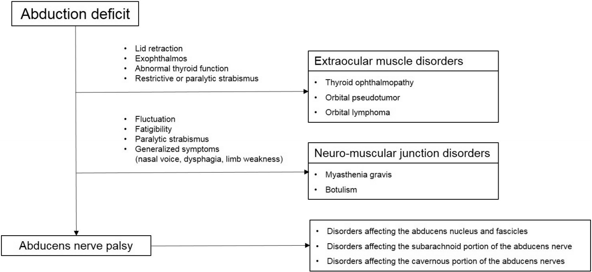
Diagnostic flow chart of abduction deficit of eye.

Etiology of abducens nerve palsy
눈운동장애에서 통증의 유무는 원인질환을 감별하는데 매우 중요하다. 특히 머리 전체의 두통과 함께 시신경유두부종이나 뇌수막자극징후가 관찰될 경우에는 뇌수막염이나 뇌압 상승의 가능성을 고려해야 한다[2]. 반면에 통증이 편측 눈 주위나 이마, 측두부에 국한되어 있을 경우에는 안와, 상안와틈새, 해면정맥동이나 안장옆 영역(parasellar region) 등으로 병변을 국소화할 수 있다[3].
본 환자의 경우 안구돌출, 결막충혈, 눈꺼풀뒤당김이 동반되지 않았고 증상의 일중 변동이 없었기 때문에 외안근과 신경근접합부 병변의 가능성은 낮으며, 외전신경핵과 신경다발 병변에 흔히 동반되는 안면마비 등의 신경학적 증상도 없어 좌측 외전신경마비로 인한 외전장애로 생각할 수 있었다. 또한 다른 신경학적 징후나 시신경유두부종, 뇌수막자극징후가 없고 통증이 편측 이마에 국한되어 있어 안와 또는 해면정맥동 부위의 병변을 우선 고려할 수 있었다.
혈액검사에서 전혈구검사, 일반화학검사, 갑상선기능검사, 적혈구 침강속도 및 C-반응단백수치는 모두 정상범위였다. 뇌 자기공명영상에서 좌측 해면정맥동이 확장되어 있었으며, 해면정맥동 내에 T1과 T2 강조영상에서 모두 등신호강도(isointense signal)로 보이는 종괴(mass)가 관찰되었다. 이 병변은 조영제 투여 후 비교적 균질(homogenous)한 조영증강을 보였다(Fig. 3-A, B). 뇌자기공명혈관조영술은 정상이었다. 류마티스인자, 보체검사(C3, C4), 항핵항체(ANA), 안지오텐신전환효소(angiotensin converting enzyme), 항인지질항체(antiphospholipid antibody), 항카디오리핀항체(anticardiolipin antibody)를 포함하는 결체조직검사는 모두 정상이었고 뇌척수액검사도 정상이었다.
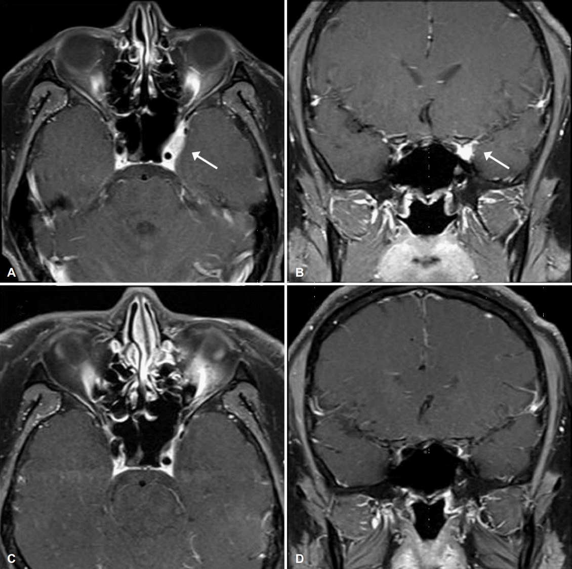
Initial gadolinium-enhanced T1-weighted MRIs (A, B) revealed a homogeneously enhancing mass lesion at the left cavernous sinus and superior orbital fissure (arrows). Follow up MRIs (C, D) performed two months after steroid treatment disclose disappearance of the enhancing mass lesion. MRI; magnetic resonance imaging.
질문 2. 감별진단은?
뇌 자기공명영상검사에서 해면정맥동 내 국소적으로 조영증강되는 종괴가 관찰될 경우 크게 감염성 질환, 염증성 질환과 종양을 감별해야 한다[3]. 감염성 질환으로는 세균이나 곰팡이, 결핵 등이 있고, 주변의 부비동염(sinusitis)이 해면정맥동으로 침윤되는 경우가 많다[3]. 염증성 질환은 사코이드증(sarcoidosis), 베게너육아종증(Wegener’s granulomatosis), 결절다발동맥염(polyarteritis nodosa), Tolosa-Hunt증후군 같은 질환이 원인이 될 수 있다[4]. 사코이드증이나 결체조직질환들은 동반되는 전신증상과 자가면역항체검사 등으로 진단할 수 있다. 종양의 경우 수막종(meningioma)이나 해면혈관종(cavernous angioma), 뇌하수체선종(pituitary adenoma), 비인두암종(nasopharyngeal carcinoma), 림프종(lymphoma) 그리고 전이성 종양 등을 감별해야 한다[3]. 이들은 대부분 주변의 안장오목(sellar fossa)이나 중두개와(middle cranial fossa)에서 해면정맥동 내로 침범하는 경우가 많다[2].
본 환자의 경우 감염성 질환을 의심할 만한 부비동염 소견은 보이지 않았으며, 뇌척수액검사, 안지오텐신전환효소와 결체조직검사는 모두 정상이었다. 따라서 해면정맥동 내 특징적인 뇌 자기공명영상 소견과 편측 눈주위 통증을 동반한 눈운동신경마비로 미루어 볼 때 Tolosa-Hunt증후군의 가능성이 매우 높다.
토 의
Tolosa-Hunt증후군은 통증안근마비(painful ophthalmoplegia)를 주 증상으로 하는 해면정맥동 내 비특이적 육아종성염증이다[5]. 모든 연령에서 발생할 수 있고, 성별에 따른 차이는 없다[3]. 주로 편측으로 발생하지만 간혹 양측으로 발생하기도 한다. 통증의 위치는 눈 주위가 가장 흔하고, 종종 눈 뒤, 이마, 그리고 측두부로 뻗칠 수 있다. 눈운동신경마비는 통증과 동시에 발생하거나 통증 발생 2주 이내에 나타난다[5,6]. 눈돌림신경(oculaomotor nerve), 도르래신경(trochlear nerve), 외전신경(abducens nerve)의 세 가지 눈운동신경을 모두 침범할 수 있고, 이들의 다양한 조합으로 함께 마비가 발생하기도 한다[5,6]. 삼차신경(trigeminal nerve)이나 시신경(optic nerve)까지 침범하여 안면감각저하나 시력저하가 발생할 수 있다[5,6].
진단은 특징적인 임상증상과 영상검사로 하지만, 통증안근마비를 일으키는 여러 가지 염증, 감염, 혈관질환과 종양 등을 배제하는 것이 중요하다(Table 2) [1]. 과거에는 스테로이드에 대한 좋은 반응을 Tolosa-Hunt증후군 진단에 중요한 기준으로 삼았지만, 치료에 대한 반응은 환자마다 다양하고 다른 질환에서도 스테로이드에 반응이 좋을 수 있기 때문에 주의를 요한다. 그리고 뇌 자기공명영상을 통하여 해면정맥동 내 병변의 확인과 다른 질환의 배제가 가능하기 때문에, 최근에 발표된 국제두통질환분류 3판(3rd edition of International Classification of Headache Disorders, ICHD-3)에서는 스테로이드에 대한 반응성을 제외하고 뇌 자기공명영상의 역할이 강조된 새로운 진단기준을 제시하였다(Table 3) [5].

Causes of painful ophthalmoplegia

International classification of headache disorders-3 diagnostic criteria for Tolosa-Hunt syndrome
뇌 자기공명영상에서 Tolosa-Hunt증후군의 전형적인 소견은 해면정맥동 내 국소 혹은 미만성의 연부조직 침윤이 관찰되고 주변 조직의 부종과 조영증강을 보이는 것이다[4]. 일부에서는 상안와틈새나 안와까지 조영증강을 보이기도 하지만 뼈의 미란은 관찰되지 않는다[4]. 하지만 임상적으로 Tolosa-Hunt증후군이 의심되더라도 뇌 자기공명영상이 정상인 경우도 있으며, 임상증상과 뇌 영상에서 보이는 병변 간에 불일치를 보일 수도 있다[4]. 또한 국제두통질환분류의 진단기준에 대한 유효성 평가 연구를 살펴보면, 임상적으로 Tolosa-Hunt증후군이 의심되는 환자들 중 진단기준에 부합하는 경우는 각각 ICHD-1에서 87.5%, ICHD-2에서 60%, ICHD-3에서 47.5%로 나타났다[7]. 이는 최근 기준이 뇌 자기공명영상 소견을 포함함으로써 엄격하게 진단에 제한을 두고 있기 때문이다. 또한 ICHD-3의 진단기준에 따르면 통증이 한쪽 눈 혹은 눈 주변에 국한되어야 하며, 눈운동신경마비는 통증과 비슷한 시기 혹은 통증 발생 2주 내에 나타나야 한다. 하지만 통증 위치가 정확하게 확인되지 않거나 통증이 발생한지 2주가 넘어 복시가 발생하는 등 비전형적인 경과를 보이는 경우도 있다[8]. 따라서 진단기준을 단계로 나누어 조정하는 등 향후 ICHD-3 진단기준에 대한 재고가 있어야 하겠다[7].
Tolosa-Hunt증후군의 병태생리가 비특이적인 육아종성염증으로 추정되기 때문에 스테로이드 치료를 우선 시도하여 볼 수 있다. 1961년 처음으로 Hunt가 Tolosa-Hunt증후군에서 스테로이드 전신 요법의 효과에 대하여 언급하였지만, 현재까지 스테로이드의 적절한 치료 용량과 사용 기간에 대해서는 알려진 바가 없다[9]. 하루에 프레드니솔론 30 mg 이하의 저용량 요법에서 메틸프레드니솔론(methylprednisolone) 1 g의 고용량 요법까지 다양한 투약 방법이 제시되고 있다[10]. 스테로이드를 투여하면 눈 주위 통증은 빠른 시간 내에 감소하지만, 동반된 눈운동신경마비는 좀 더 천천히 호전된다[8]. 또한 스테로이드는 림프종이나 수막종에서도 일시적으로 효과가 있기 때문에 스테로이드의 효과 판정에 주의를 요한다. 일반적으로 수주 혹은 수개월에 걸쳐 서서히 감량하지만, 일부에서는 감량 도중 재발하기도 한다. 재발이 잦은 경우에는 스테로이드를 줄이면서 면역억제제의 투여가 도움이 될 수 있다[10].
KEY POINTS
1. 통증안근마비의 감별진단
• 혈관성: 경동맥해면정맥동루(carotid-cavernous fistula), 해면정맥동혈전증(carotid sinus thrombosis), 내경동맥류(internal carotid aneurysm)
• 염증성: Tolosa-Hunt증후군, 사코이드증(sarcoidosis), 베게너육아종증(Wegener’s granulomatosis), 결절다발동맥염(Polyarteritis nodosa), 세균, 곰팡이, 결핵 등의 감염성질환
• 종양성: 수막종(meningioma), 뇌하수체선종(pituitary adenoma), 해면혈관종(cavernous angioma), 비인두암종(nasopharyngeal carcinoma), 림프종(lymphoma)
• 기타: 당뇨병안근마비(diabetic ophthalmoglegia), 편두통안근마비(ophthalmoplegic migraine), 거대세포동맥염(giant cell arteritis)
2. 통증안근마비에서 Tolosa-Hunt증후군으로 진단을 위해서는 임상증상과 뇌 자기공명영상에서 육아종성종괴 소견 확인 및 다른 질환의 배제가 필요하며, 진단기준에 맞지 않더라도 임상적으로 의심되면 추적검사 및 스테로이드 사용이 권고된다.
3. Tolosa-Hunt증후군은 저절로 호전되는 질환으로 알려져 있지만, 잦은 재발과 영구적인 뇌신경마비를 유발할 수 있기 때문에 적절한 진단과 치료가 필요하다.