초급성 허혈뇌졸중 환자에서 정맥내혈전용해 치료 이후 나타난 고신호강도급성재관류표지자
Hyperintense Acute Reperfusion Marker after Intravenous Thrombolysisin a Patient with Hyperacute Ischemic Stroke
Article information
69세 남자가 3시간 전 시작한 우측 위약감과 언어장애로 내원하여, 신경학적 진찰에서 NIH 뇌졸중척도(National Institutes of Health Stroke Scale, NIHSS)는 5점이었다. 두개내외혈관의 협착은 없었으나(Fig. A), 확산강조영상에서 좌측의 급성기 허혈병변들이 관찰되었다(Fig. B). 재조합조직플라스미노겐활성제(recombinant tissue plasminogen activator, r-tPA) 투여 완료 3시간 이후 생체활력징후 등은 정상이었으나, 의식저하 및 우측 반신의 완전마비가 발생하였다(NIHSS 12점). 증상 악화 3시간째 액체감쇠역전회복영상, T1 조영증강영상, 기울기에코영상에서 좌측 대뇌 반구 피질을 따라 고랑과 수조의 고신호 음영이 관찰되었다(Fig. C). 고신호강도급성재관류표지자(hyperintense acute reperfusion marker, HARM)로 진단하여, 두개내압 및 혈압을 조절하였다[1,2]. 사흘만에 의식을 포함하여 우측마비와 언어장애가 호전되었다. r-tPA혈전용해치료 이후 급격한 임상적 악화와 영상검사에서 재관류손상으로 인한 HARM을 확인하여 이를 보고하고자 한다.
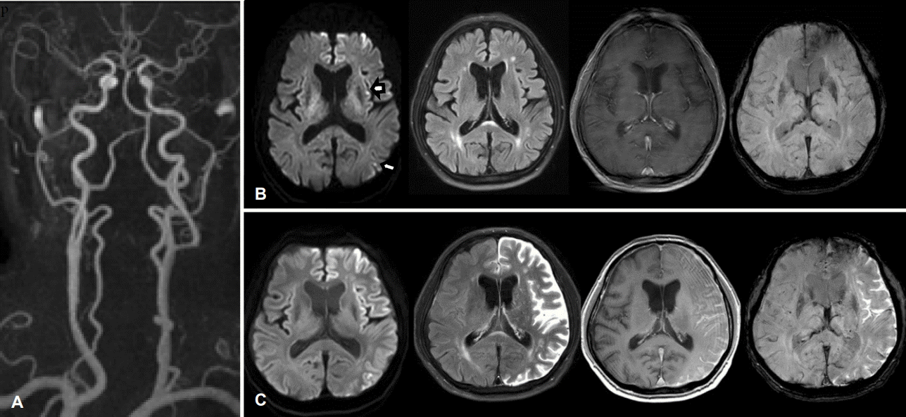
Initial MRA, MRI (A, B) and follow up MRI (C) after recombinant tissue plasminogen activator (r-tPA) infusion. (A) Enhanced MRA showed no definite steno-occlusive lesions during IV r-tPA infusion. (B) Initial DWI shows multiple high signal lesions on the left cerebral hemisphere (arrows). (C) Brain MRI, performed 6 hours after IV r-TPA infusion revealed newly appeared hyperintense signal within the whole sulci and cisterns of the left hemisphere on T2 FLAIR, T1-enhanced and GRE images. MRA; magnetic resonance angiography, MRI; magnetic resonance imaging, DWI; diffusion-weighted image, FLAIR; fluid-attenuation inversion recovery, GRE; gradient echo image.