원인불명 색전뇌경색 환자에서 폐동정맥루 단순 흉부 방사선 사진 소견
Simple Chest Radiography Findings of Pulmonary Arteriovenous Fistula in Patients with Otherwise Cryptogenic Ischemic Stroke
Article information
모순색전(paradoxical embolic)뇌경색으로 방문한 두 명의 환자(46세, 56세 여자 환자)가 단순 흉부방사선사진에서 이상을 보여, 흉부전산단층혈관촬영을 통해 폐동정맥루(pulmonary arteriovenous fistula)를 진단하였다. 두 환자의 단순흉부방사선사진에서는 둥근 타원형 모양의 경계가 분명하고, 균질한 혼탁의 병변을 보였다(Fig. 1, 2). 이 병변은 조영증강 흉부전산단층혈관촬영을 통해 폐동정맥루로 확진되었다. 원인불명의 색전뇌경색에서 폐동정맥루는 색전 원인의 중요한 감별진단 중에 하나이다. 이를 위한 선별검사인 단순흉부 방사선사진에서는, 일반적으로 연조직 음영의 종괴로 보이나, 주변의 혈관음영과는 다른 방향성을 보인다[1].

46-year-old woman with embolic infarction. Chest X-rays (A) revealed round mass with sharply defined border (red arrow) which were confirmed as PAVM by contrast-enhanced chest CT (B). Diffusion weighted images demonstrated ischemic lesion in left angular gyrus (C). PAVM; pulmonary arteriovenous malformation, CT; computed tomography
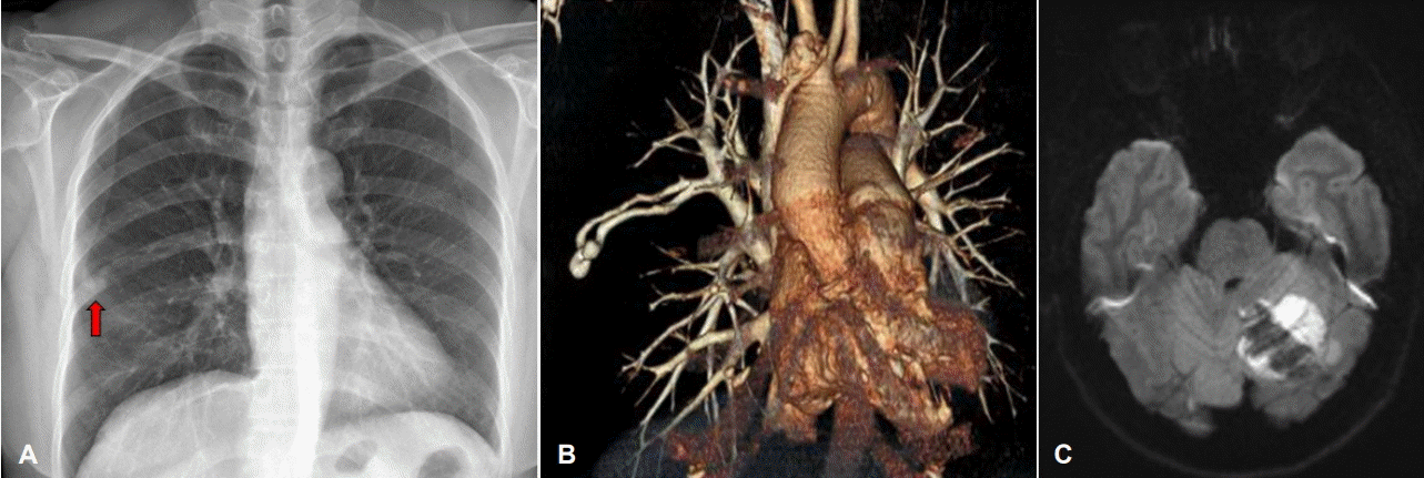
56-year-old woman with embolic infarction. Chest X-rays (A) demonstrated ovoid convoluted mass with demarcated border (red arrow) which were diagnosed as PAVM by contrast-enhanced chest CT angiography (B). Diffusion weighted images demonstrated ischemic lesion with hemorrhagic transformation in left cerebellum (C). PAVM; pulmonary arteriovenous malformation, CT; computed tomography.