두개내 유피낭종 파열에 의한 벼락두통

Thunderclap Headache Due to Ruptured Intracranial Dermoid Cyst

Article information

J Korean Neurol Assoc. 2018;36(2):134-135
Publication date (electronic) : April 30, 2018
doi : http://dx.doi.org/10.17340/jkna.2018.2.19
Department of Neurology, Gachon University Gil Medical Center, Incheon, Korea
김현석, 신동훈, 최석홍
가천대학교 길병원 신경과
Address for correspondence: Dong Hoon Shin, MD Department of Neurology, Gachon University Gil Medical Center, 21 Namdong-daero 774beon-gil, Namdong-gu, Incheon 21565, Korea Tel: +82-32-460-3346 Fax: +82-32-460-3344 E-mail: dr.donghoon.shin@gmail.com
received : November 20, 2017 , rev-recd : January 25, 2018 , accepted : January 25, 2018 .

고혈압이 있는 47세 여자가 10일 전부터 시작한 벼락두통으로 내원하였다. 두통은 시각아날로그척도 8점으로 양측 관자놀이에서 시작하여 이마로 퍼지는 양상이었고 구역과 가슴떨림이 동반되었다. 신경학적 진찰에서 뇌막자극징후 외에 다른 이상은 없었다. 혈액검사에서 백혈구 수치 9.55×10³/mm로 경도의 백혈구증가증이 있었고, C-반응단백질과 적혈구침강비율은 정상이었다. 뇌척수액검사는 정상이었다. 뇌 컴퓨터단층촬영에서 좌측 소뇌다리뇌수조(cerebellopontine cistern)로 지름 5 mm 지질성 병변이 있었고(Fig. A), 뇌 자기공명영상에서 대뇌겸(falx cerebri)과 천막(tentorium)에 두개내 유피낭종 파열이 의심되는 신호변화들이 관찰되었다(Fig. B-H). 이에 잘토프로펜, 니모디핀, 프로프라놀롤을 투여하였고, 두통은 시각아날로그척도 5점으로 호전되었다. 두개내 유피낭종은 대부분 무증상이지만 크기가 커지면 종괴 효과가 생기거나 낭종이 파열될 경우 신경학 증상을 보이기도 하며, 벼락두통 양상으로 증상이 나타나기도 한다[1,2]. 저자들은 두개내 유피낭종 파열로 인한 벼락두통을 경험하여 이를 보고하는 바이다.
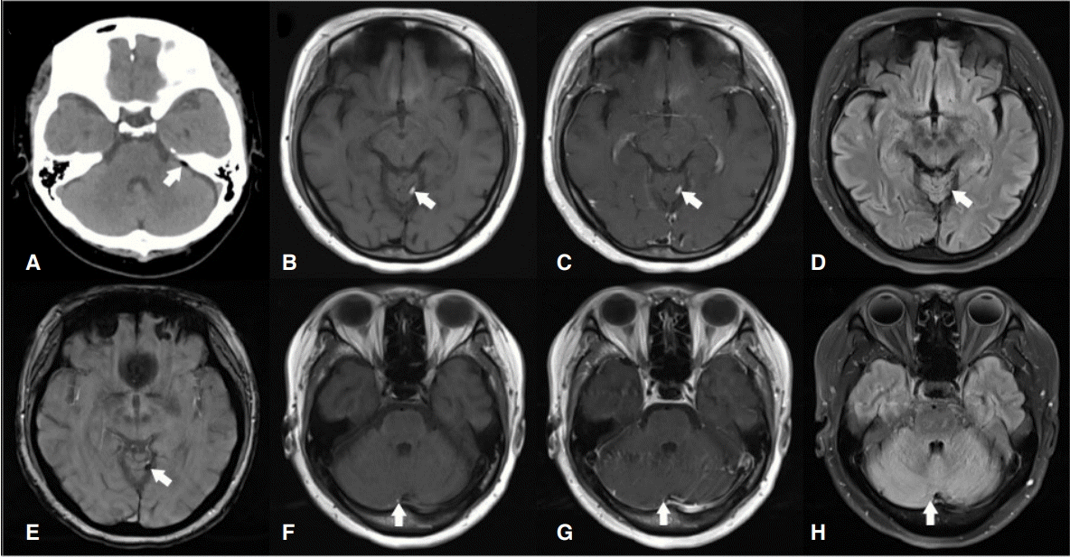
Figure.

Image findings of dermoid cystic rupture. Non-enhance computed tomography scans showed a hypodense focal fatty lesion in the left cerebellopontine angle cistern (A). Hyperintense on T1-weighted image (B and F), blooming artifact on gradient echo sequences (E), sequence suppression on fat suppression fluid attenuated inversion recovery (D and H), and non-enhancing lesions on T1 contrast enhance image (C and G) were noted in falx and tentorium, which means fat signal. It can be assumed as dermoid cystic rupture.

References

1. Şalçini C, Hatıloğlu D, Evrensel A, Tanrıdağ AO. Episodic headache due to ruptured intracranial dermoid cyst. BMJ Case Rep 2015;2015pii: bcr2015209691.
2. Ray MJ, Barnett DW, Snipes GJ, Layton KF, Opatowsky MJ. Ruptured intracranial dermoid cyst. Proc (Bayl Univ Med Cent) 2012;25:23–25.

Article information Continued

Figure.

Image findings of dermoid cystic rupture. Non-enhance computed tomography scans showed a hypodense focal fatty lesion in the left cerebellopontine angle cistern (A). Hyperintense on T1-weighted image (B and F), blooming artifact on gradient echo sequences (E), sequence suppression on fat suppression fluid attenuated inversion recovery (D and H), and non-enhancing lesions on T1 contrast enhance image (C and G) were noted in falx and tentorium, which means fat signal. It can be assumed as dermoid cystic rupture.