철결핍빈혈을 동반한 대동맥궁 혈전으로 유발된 색전뇌경색
Cerebral Embolic Infarction Caused by Aortic Arch Thrombus with Iron Deficiency Anemia
Article information
Trans Abstract
Atherosclerotic lesions at the aortic arch are recognized as critical sources of embolic strokes. However, there have been few case reports of aortic arch thrombus occurring without atherosclerotic changes, especially those related to iron deficiency anemia (IDA). A 44-year-old woman was admitted due to rapid-onset right hemiparesis and aphasia. Etiological investigations for cerebral infarction revealed no abnormality other than IDA. This is a rare case of cerebral embolic infarction caused by an aortic arch thrombus with IDA in a middle-aged woman.
대동맥궁의 죽상경화는 색전뇌경색의 중요 원인으로 알려져 있지만, 죽상경화 없이 혈전으로 인한 색전뇌경색이 오는 경우는 흔하지 않다[1]. 대동맥궁 혈전을 보인 대규모 연구에 의하면 아주 소수의 환자들에서만(0.08%) 죽상경화 없이 대동맥궁 혈전을 보였다[2]. 죽상경화로 인한 색전뇌경색을 보인 환자들은 고령이었으며[3], 반대로 죽상경화 없이 발생한 뇌경색 환자들은 상대적으로 젊었다[2]. 대동맥궁의 혈전을 유발하는 원인은 고혈소판증, 적혈구증가증, C단백질결핍(protein C deficiency), 철결핍빈혈(iron deficiency anemia, IDA)과 같은 요인들이 있다. IDA로 인한 대동맥궁의 혈전으로 발생된 색전뇌경색 환자들의 경우 젊은 여성이 많았으며, 시간이 지나면서 대동맥궁의 혈전은 관해되는 모습을 보였다[4,5]. 하지만 빈혈로 인해 대동맥궁의 혈전이 생기는 경우는 매우 드물고 아직까지 국내에서 보고된 바는 없다. 저자들은 젊은 여성에서 IDA로 인한 대동맥궁의 혈전으로 색전뇌경색이 발생한 예를 경험하여 문헌고찰과 함께 이를 보고하고자 한다.
증 례
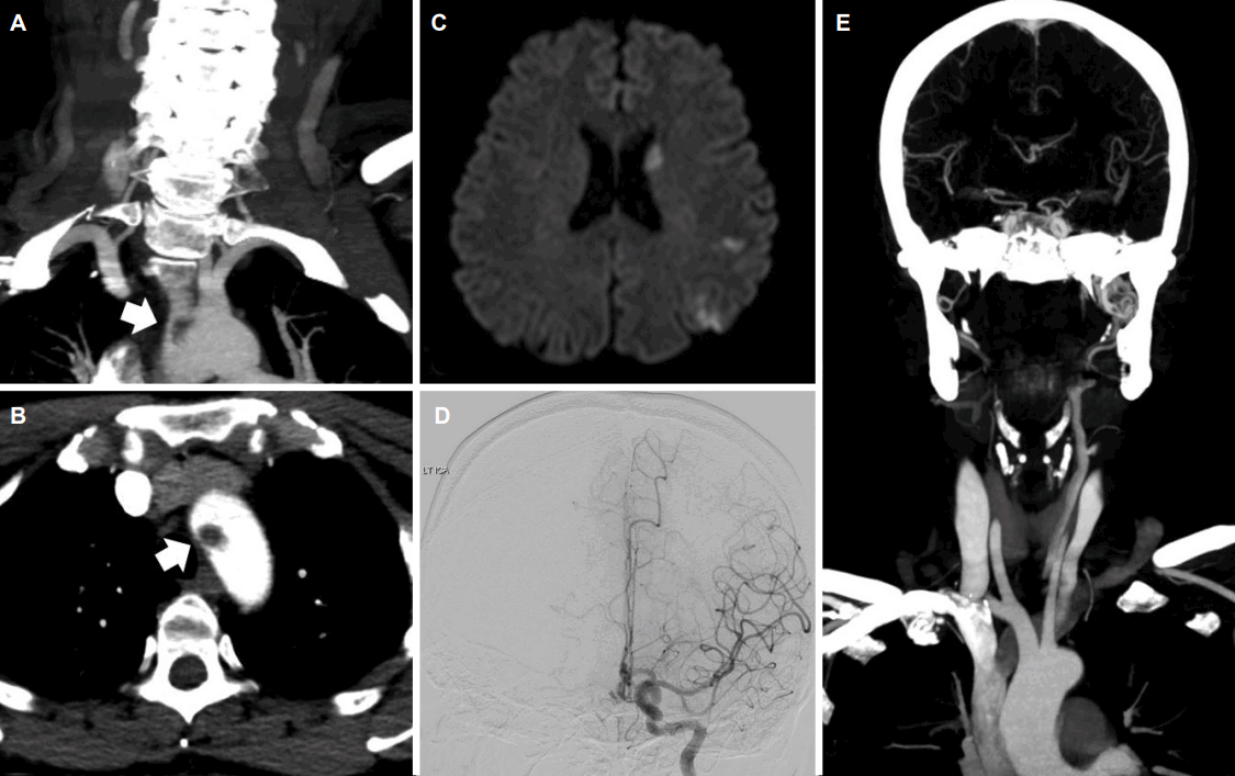
44세 오른손잡이 여자가 갑자기 발생한 우측 팔다리의 반신마비 및 완전실어증을 주소로 응급실을 내원하였다. 환자는 15년 전 자궁근종을 진단받았으며 이로 인한 월경과다증이 있는 것과 흡연 외에 다른 과거력은 없었다. 방문 당시 혈압은 160/100 mmHg이었고 맥박은 82회/분, 호흡수는 19회/분 체온은 37.2℃였다. 신경학적진찰에서 의식은 명료하였으며 우측 팔다리의 반신마비와 완전실어증, 우측 중추성 얼굴마비, 우측 반맹 소견을 보였다. 심전도검사에서 특이 소견은 보이지 않았다. 뇌컴퓨터단층촬영(computed tomography, CT)에서 급성뇌출혈 및 뇌경색 소견은 보이지 않았다. CT혈관조영술에서 내경동맥 및 대뇌동맥의 협착이나 폐색은 보이지 않았으나, 대동맥궁에서 혈전이 관찰되었다(Fig. A, B). 디지털감산혈관조영술(digital subtraction angiography)에서 왼쪽 내경동맥 및 대뇌혈관의 협착 및 폐색은 보이지 않았다(Fig. D). 증상 발생 2시간째 시행한 뇌자기공명영상(magnetic resonance image, MRI)의 확산강조영상(diffusion weighted image, DWI)에서 다발성의 작은 병변들이 왼쪽 대뇌피질과 백질 부위에 보였다(Fig. C). 혈액검사에서 혈색소는 7.2 g/dL, 적혈구용적률(hemotocrit)은 28.9%, 백혈구는 8,580/mm3 , 혈소판은 400,000/mm3 이었고 평균적혈구용적(mean corpuscular volume) 64 fL, 평균적혈구혈색소(mean corpuscular hemoglobin) 16 pg이었다. 페리틴(ferritin) 6.53 ng/mL, 혈청철 17.8 μg/dL, 트랜스페린(transferrin) 2.26 mg/dL을 보였다. C단백질 활성(protein C activity) 60%, S단백질활성(protein S activity) 40%로 감소된 소견을 보였으며, 그 외 항트롬빈III(antithrombin Ⅲ), 항카디오리핀항체(anticardiolipin antibody), 항인지질항체(antiphospholipid antibody), 루푸스항응고인자(lupus anticoagulant), 호모시스테인(homocysteine) 결과는 모두 이상 없었다. IDA의 원인을 찾기 위한 추가적인 검사를 하였으나 자궁근종 외의 다른 원인은 발견할 수 없었다.

Brain MRI and angiography of patient. (A) Sagittal image on the neck CT angiography showed thrombus (7.4 x 10.6 mm) in aortic arch (arrow). (B) Axial image on the neck CT angiography showed same thrombus (arrow). (C) Diffusion weigted MRI showed multiple acute infarction in the left MCA territory. (D) Digital subtraction angiography showed no stenoocclusive lesion from the left internal carotid artery to MCA. (E) Follow up neck CT angiography after 1 week, thrombus was not seen in aortic arch. CT; computed tomography, MRI; magnetic resonance image, MCA; middle cerebral artery.
환자의 IDA는 수혈 이후 호전되었으며, 내원일부터 실로스타졸(cilostazol) 200 mg을 복용하였다. 증상 발생 48시간 후부터 환자는 보행기를 잡고 보행을 할 수 있을 정도로 호전되었으며 경미한 구음장애를 보였다. 증상 호전 이후 7일째 시행한 뇌CT혈관조영술에서 대동맥궁에 위치하던 혈전은 관찰되지 않았다(Fig. E). 같은 날 시행한 식도경유심장초음파(transesophageal echocardiography)에서도 대동맥궁 혈전은 보이지 않았으며 열린타원구멍(patent foramen ovale)은 없었다. 이후 환자는 증상이 더욱 호전되어 현재 외래에서 추적관찰 중이며 후유증은 거의 없는 상태이다.
고 찰
본 증례의 환자는 죽상경화와 같은 흔한 병인없이 대동맥궁의 혈전이 CT혈관조영술에서 확인 되었다. 또한 심각한 IDA 소견을 보였으며 이로 인해 응고항진상태(hypercoagulability state)가 유발되고, 대동맥궁에 혈전이 발생되었을 것으로 생각한다[6]. 그리고 이렇게 생성된 혈전에서 떨어져 나간 작은 혈전으로 인해 색전뇌경색이 발생했다고 볼 수 있다.
저자들의 보고와 유사하게 젊은 여성에서 IDA로 인한 대동맥궁의 혈전으로 인해 색전뇌경색이 발생한 경우가 있었다[4,5]. 이 경우 흡연을 제외한 다른 심혈관계 위험인자는 없었으며, 대동맥 혹은 대뇌동맥의 협착이나 폐색은 보이지 않았다. 또한, 나이가 비교적 젊은 여성들에서 월경과다증으로 인한 IDA를 동반하는 경우가 많았고, 일부에서는 자궁근종 혹은 자궁섬유화(uterine fibroids)가 월경과다증의 원인인 경우가 있었다. 대동맥궁의 혈전은 식도경유심장초음파를 통해 확인하는 것이 일반적이지만, 본 증례의 경우 뇌경색과 관련된 혈관 이상을 확인하기 위해 시행한 CT혈관조영술에서 대동맥궁의 혈전을 확인할 수 있었다. 이는 경동맥이나 대뇌동맥 폐색이 의심되는 급성뇌경색에서 대동맥궁을 포함한 CT혈관조영술이 색전뇌경색의 원인을 찾는데 도움이 될 수 있다는 것을 보여주는 예라고 하겠다.
지금까지 알려진 바로는 대동맥의 죽상경화로 인해 발생한 혈전이 주로 색전뇌경색의 중요한 원인으로 알려져 있다[1]. 하지만 저자들의 증례와 다른 보고들을 비추어 볼 때 심각한 IDA가 응고항진상태를 야기함으로 인해 대동맥궁 혈전이 생겼다고 생각된다. 이러한 혈전 형성에 대해 몇 가지 가설을 생각해 볼 수 있는데, 크게 두 가지 기전으로 나누어 볼 수 있다. 첫 번째는 전신적인 과응고상태가 유발되어 혈전 형성이 일어날 수 있다는 가설 중 우선 급성출혈로 인한 빈혈은 혈소판증가증(thrombocytosis) 및 혈소판부착성(platelet adhesiveness) 증가를 가져올 수 있고[7], 이로 인하여 혈전생성(thrombogenesis)이 촉진될 수 있다는 것이다. 다른 가설로는 빈혈로 인해 플라스미노겐활성억제인자-1(plasminogen activation inhibitor-1)이 증가되며, 이로 인해 섬유소용해능(fibrinolytic activity)이 감소됨으로 혈전형성이 일어날 수 있다는 것이다[8]. 두 번째는 대동맥궁에서 빈혈 자체가 혈역학장애(hemodynamic disturbance)를 유발하며 전단응력(shear stress) 증가로 내피부착분자유전자(endothelial adhesion molecule genes)의 상향조절(upregulation)이 유발됨으로써 혈전이 형성된다는 것이다[9]. 이로 인하여 면역학적 염증반응(immunologic-inflammatory reaction)으로 인해 혈전생성이 일어날 수 있다고 생각된다. 본 증례의 경우 반응혈소판증가증(reactive thrombocytosis)은 보이지 않았으나, S단백질활성이 감소되어 응고항진상태가 유발된 것으로 판단할 수 있었다. 저자들의 증례 및 다른 보고들의 혈전들은 모두 대동맥궁에 생겼다. 동맥관은 태생 이후 수개월 이내에 막히며 이후 동맥관인대(ligamentum arteriosum)로 대동맥궁 아래에 위치하게 되는데, 동맥관인대 부착 부위의 석회화는 성인에게 흔히 발견되고, 나이가 들수록, 죽상경화가 있을수록 발견 빈도는 높았다[10]. 따라서, 대동맥궁이 대동맥의 다른 부위에 비해 혈전 생성이 더 잘 발생할 수 있다고 생각되지만, 본 증례의 경우 동맥관인대 부착 부위의 석회화는 보이지 않았다. 향후 대동맥의 다른 부위에 비해 대동맥궁에 혈전이 잘 생기는지 여부에 대한 대규모의 임상 연구가 필요하다고 생각된다.
결론적으로, 심각한 빈혈은 대동맥궁 혈전 생성에 중요한 원인이 될 수가 있다. 따라서 잠재뇌졸중(cryptogenic storke) 환자에서 IDA가 동반된 경우 대동맥궁 혈전을 찾는 것을 고려해야 하며, 특히 젊은 여성에서 IDA가 있는 경우 자궁근종 등의 출혈 원인을 찾아 재출혈을 방지하는 것이 중요할 것으로 생각된다.
Acknowledgements
이 논문은 2017년도 조선대학교병원 선택진료 학술연구비에 의하여 연구되었음.