소뇌경색으로 발현된 후하소뇌동맥류내 자발혈전증
Spontaneous Thrombosis of Posterior Inferior Cerebellar Artery Aneurysm Presenting as Cerebellar Infarction
Article information
Trans Abstract
Ischemic stroke caused by spontaneous thrombosis of posterior inferior cerebellar artery (PICA) aneurysm has been rarely reported. A 52-year-old man presented with sudden headache, dizziness, and gait disturbance. Diffusion-weighted MRI showed acute infarction in left PICA territory. A saccular aneurysm with internal thrombus at the distal PICA was detected by CT angiography and conventional angiography. The thrombus resolved spontaneously at 2 months after stroke onset with aspirin medication. At that time, endovascular coiling was underwent successfully to prevent aneurysmal rupture.
뇌동맥류(cerebral aneurysm)는 주로 거미막하출혈(subarachnoid hemorrhage)을 일으키는 질환으로 뇌내동맥류 중에 후하소뇌동맥류(posterior inferior cerebellar artery aneurysm)의 빈도는 적다고 알려져 있으며 후하소뇌동맥의 원위부 부분에 발생하는 경우는 더욱 더 드물다고 알려져 있다[1]. 뇌동맥류에서 때때로 허혈증상이 나타나기도 하며 그중에는 자발혈전증(spontaneous thrombosis)에 의한 경우가 있다[2]. 저자들은 원위부 후하소뇌동맥류내 자발혈전증에 의한 소뇌경색의 예를 경험하여 보고하고자 한다.
증 례
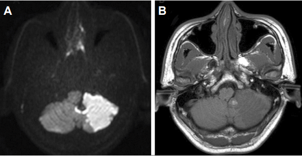
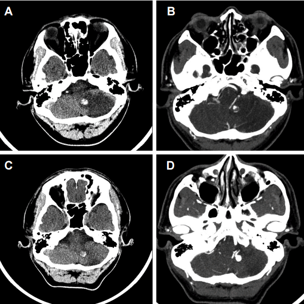
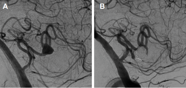
52세 남자가 운전 도중에 갑자기 발생된 심한 두통과 어지럼으로 응급실로 내원하였다. 환자는 걸을 때 왼쪽으로 기우는 증상이 수분 동안 지속된 후 호전되었으나 두통과 어지럼이 수일간 지속되어 응급실에 왔다. 환자의 내원 당시 활력징후(vital sign)는 수축기혈압/이완기혈압 123/70 mmHg, 맥박 89회/분, 체온 36.9도 였다. 환자는 고혈압, 당뇨병, 심혈관 질환은 없었으며 흡연은 하지 않았다. 가족력으로 아버지의 심근경색 병력이 있었다. 응급실에서 시행한 신경학적진찰에서는 국소신경학적 결손은 관찰되지 않았고 환자는 두통과 어지럼만을 호소하였다. 확산강조영상(diffusion-weighted image)에서 좌측 후하소뇌동맥 영역에 고신호강도를 보이고 있으며(Fig. 1-A) 뇌자기공명혈관조영술(brain magnetic resonance angiography)에서는 좌측 후하소뇌동맥 원위부에서 소낭동맥류(saccular aneurysm)가 관찰되었다. 이외에 두개내 및 두개외 다른 혈관의 협착이나 동맥류는 발견되지 않았다. 또한 T1강조영상(T1 weighted MRI) 및 뇌전산화단층혈관조영술(brain CT angiography)에서 동맥류내 혈전으로 생각되는 원형 모양의 비균질 병변이 좌측 소뇌 내측부와 연수 사이에서 관찰되었고 이는 동맥류내 공간을 부분적으로 차지하고 있었다(Fig. 1-B, 2-A, B). 고식적뇌혈관조영술(conventional angiography)을 시행하였고 좌측 후하소뇌동맥의 편도 부분(tonsilar segment)의 동맥류를 확인하였다(Fig. 3-A). 처음 시행한 심전도와 2개월 후 다시 시행한 심전도는 정상 리듬이었고 두개경유도플러(transcranial Doppler)검사에서 미세색전(microembolism) 소견은 관찰되지 않았다. 흉부경유심초음파검사(transthoracic echocardiography)는 시행하지 않았다. 항인지질증후군(antiphospholipid syndrome) 및 혈액응고장애를 평가하기 위해 시행한 혈액검사(anticardiolipin antibody, anti beta 2 glycoprotein I antibody, lupus anticoagulant, D-dimer, antithrombin III activity, protein C/S activity, protein C/S antigen)에서는 특이소견이 없었다. 이에 저자들은 환자가 다른 뇌경색의 위험인자를 가지지 않았고 뇌영상에서 다른 뇌혈관의 이상이나 뇌실질의 이상을 찾을 수 없었으며 심장성색전증의 증거가 없었기 때문에 좌측 후하소뇌동맥 원위부 소낭동맥류에 있는 동맥류내 혈전에 의한 색전성 소뇌경색으로 진단하였다. 이에 아스피린(aspirin; Aspirin Protect Tab. 100mg, Bayer AG, Seoul, Korea) 300 mg을 투여하였으며 환자는 입원 중 신경학적 이상은 없었고 두통 및 어지럼은 호전되어 퇴원하였다. 뇌경색 발생 2달 후에 재시행한 뇌전산화단층혈관조영술에서는 동맥류내 혈전이 사라진 것을 확인하였다(Fig. 2-C, D). 환자는 간헐적인 두통 외에 다른 신경학적 이상은 없었으나 동맥류의 크기(9 × 6.6 × 5.8 mm)가 컸기 때문에 뇌동맥류 파열의 위험이 있어 혈관내중재술(endovascular intervention)을 시행하였다(Fig. 3-B). 환자는 시술 이후에 별다른 합병증은 없었으며 아스피린 100 mg을 복용하면서 외래에서 추적 관찰 중이다.

Initial MRI finding. MRI shows acute infarction in the left cerebellum in diffusion-weight image (A) and a large aneurysm with partial internal thrombus of the left PICA in T1 weighted MRI (B). MRI; magnetic resonance imaging, PICA; posterior inferior cerebellar artery.

Initial and follow-up CT and CT angiography. Initial CT image shows infarction in the left cerebellum and a large aneurysm with partial internal thrombus of the left PICA in non-contrast CT (A) and CT angiography (B). Internal thrombus was disappeared in follow-up CT at 2 month later (C, D). PICA; posterior inferior cerebellar artery, CT; computerized tomography.
고 찰
동맥류내의 자발혈전증은 특히 거대동맥류(giant aneurysm)에서 흔하게 관찰되는데 거대동맥류의 약 50%에서 관찰되는 것으로 보고되고 있다[3]. 거대동맥류에서 혈전이 흔하게 발생되는 것은 동맥류의 부피와 동맥류 목의 비율과 연관성이 있는 것으로 알려져 있는데, 동맥류의 크기에 비하여 목이 좁은 경우에 동맥류내에 혈전이 형성될 가능성이 높아진다[4]. 그 외에도 모혈관(parent artery)의 혈류역학, 동맥류내의 난류(turbulent flow)에 의한 혈관내피손상, 동맥류의 나이, 혈액응고장애 등이 동맥류내 자발혈전증의 형성과 관계가 있는 것으로 제시되고 있다[2].
비거대소낭동맥류(non-giant saccular aneurysm)에서는 동맥류내 혈전이 매우 드물게 형성되는 것으로 알려져 있는데 특이한 점은 동맥류의 자발혈전증에 의하여 뇌경색이나 일과성허헐발작 같은 허혈증상이 발생되는 경우는 오히려 거대동맥류보다는 비거대동맥류에서 자주 보고되고 있다는 점이다[2,5]. 동맥류내 혈전에 의하여 허혈증상이 발생되는 기전으로는 동맥류내 혈전의 이전(dislodgement)에 의한 이차적인 원위부동맥색전증(distal artery embolism), 동맥류내 혈전이 국소적으로 진행되어 모혈관폐색(parent artery occlusion)을 일으키는 기전, 덩어리효과(mass effect)에 의한 동맥압박(arterial compression)에 의한 허혈 등이 제시되고 있다. 본 증례의 경우에는 원위부동맥색전증에 의하여 발생된 뇌경색으로 판단되며 기존 문헌보고에서도 이에 의한 허혈증상이 주된 기전으로 제시되어 있다[5].
후하소뇌동맥의 동맥류는 드물게 보고되고 있으며 전체 두개내 동맥류(intracranial aneurysm)의 0.49-3.0%에 해당한다고 알려져 있다[1]. 원위부 후하소뇌동맥(distal posterior inferior cerebellar artery)에서 발생하는 동맥류는 0.28-1.4%로 더욱 드문 것으로 보고되어 있다[1].
후하소뇌동맥의 동맥류의 증상은 주로 거미막하출혈이지만 드물게 동맥류내 혈전이 관찰되는 경우가 있다[1,6-8]. 이 경우에 혈전화된 거대동맥류에 의한 덩어리효과(mass effect)로 발현되는 경우가 드물게 보고되어 있으며[1,6-8] 동맥류내 혈전에 의해 소뇌경색이 발생한 경우는 소뇌편도(cerebellar tonsil)에 작은 소뇌경색이 발생한 경우는[2] 있었으나 혈관영역성 소뇌경색(territorial cerebellar infarction)이 발생한 경우는 현재까지 보고된 예가 없는 것으로 보인다. 한 연구에서는 허혈증상을 보인 비파열뇌동맥류(unruptured aneurysm)의 경우, 주로 내경동맥(internal carotid artery)이나 중대뇌동맥(middle cerebral artery) 동맥류에서 발생하는 경우가 가장 많다고 기술하고 있으며 이 연구에서 후하소뇌동맥의 동맥류에서 허혈증상을 보인 경우는 없었다[9]. 또한 비교적 작은 동맥류(직경 10 mm 이하)의 경우 약물치료만으로 뇌경색 재발을 방지할 수 있다고 보고되고 있으며[9] 본 증례에 있어서도 출혈합병증 방지를 위한 혈관내중재술을 시행했으나 아스피린 투여 후 동맥류내 혈전은 제거된 것을 확인할 수 있었다.
동맥류내 자발혈전증 자연경과에 대해서는 정확히 알려져 있지는 않으나 기존의 보고에서 자연재개통(spontaneous recanalization)이 되는 경우가 많이 있다고 알려져 있는데 Calviere 등[5]에 의하면 뇌허혈증으로 내원한 뇌동맥류 중에서 동맥류내 혈전이 관찰된 10명의 환자 중 7명에서 부분 혹은 완전 재개통이 되었다는 연구 결과로 볼 때 자연재개통이 되는 경우가 상당히 많음을 추정할 수 있다. 본 증례에서도 항혈소판제를 투여 후 추적 영상검사 시 혈전이 제거된 것이 확인되었는데 항혈소판제에 의한 효과의 가능성보다는 자연적으로 재개통되었을 가능성이 높아 보인다.
동맥류내 혈전과 연관된 뇌허혈증상의 치료에 대해서는 아직까지 정립되어 있지 않다. 그러나 추적관찰 시 많은 경우에서 혈전이 자연적으로 재개통되는 것으로 볼 때 뇌경색이 발생된 급성기 혹은 아급성기의 수술치료보다는 약물치료를 시행하면서 주기적인 추적관찰을 하며 혈전의 소실 혹은 감소 여부에 따라서 수술치료 혹은 중재시술을 시도해 보는 것이 고려될 수 있을 것으로 보인다. 이에 대해서는 향후 전향적인 연구가 필요할 것으로 사료된다.
