비대된 발꿈치뼈 비골결절에 의한 장딴지신경 포착
Sural Nerve Entrapment by Hypertrophic Peroneal Tubercle of the Calcaneus
Article information
22세 남자가 한달 전부터 왼쪽 발등 외측이 저려 왔다. 겉보기에 왼쪽 발꿈치뼈의 비골결절이 돌출되어 있었고(Fig. A) 이 부위를 자극했을 때 Tinel징후가 유발되었지만 결절 주변의 압통이나 보행시 통증은 없다고 하였다.
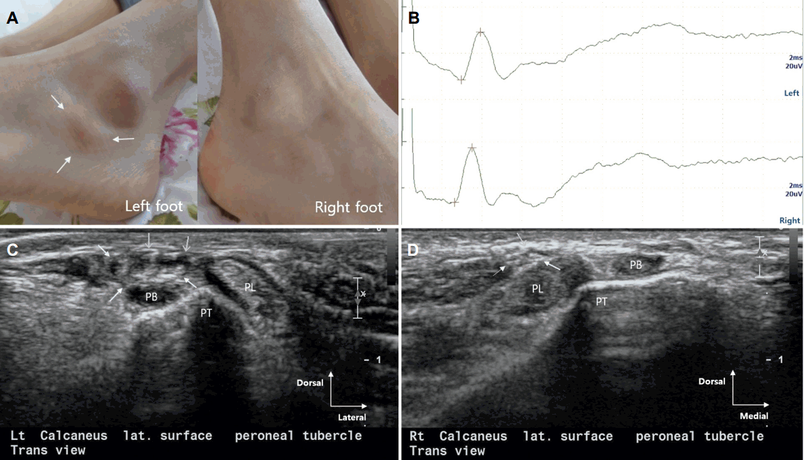
Photographs, nerve conduction studies and ultrasonogrphic images of the patient. (A) Photographs of the patient’s feet. Only in patient’s left foot, prominent peroneal tubercle is seen on the lateral aspect of the calcaneus (white arrows). (B) Sural sensory nerve conduction studies show relatively slow nerve conduction velocity of affected left sural nerve. Top trace; left sural nerve, bottom trace; right sural nerve. (C) Transverse ultrasonographic view of dorsolateral aspect of the patient’s left foot at the level of peroneal tubercle. Ultrasonographic image shows swelling and internal hypoechogenicity of the left sural nerve located between the skin and the tendon of peroneus brevis muscle (white arrows). (D) Ultrasonographic image of right sural nerve at the same aspect and level of (B). Diameter of right sural nerve is much smaller than affected left sural nerve, and honeycomb-like normal internal echotexture is preserved (white arrows). PB; tendon of peroneus brevis muscle, PT; peroneal tubercle, PL; tendon of peroneus longus muscle.
장딴지신경의 신경전도검사에서 감각신경활동전위의 진폭은 왼쪽 20.2 uV, 오른쪽 23.7 uV로 비슷하였지만 신경전달속도는 왼쪽 46.4 m/s, 오른쪽 54.7 m/s로 환측에서 상대적으로 느려져 있었다(Fig. B). 신경초음파에서는 비대된 비골결절 위에서 장딴지신경이 피부와 짧은종아리근힘줄 사이에서 부어 있었으나 종아리근힘줄들의 모양은 정상이었다(Fig. C, D).
임상적으로 비대된 비골결절은 긴종아리근힘줄을 자극하여 협착윤활막염을 종종 유발하지만[1] 장딴지신경만을 포착하는 경우는 드물다[2].
포착신경병증이 의심되나 전기생리검사가 정상이거나 이상의 정도가 확정적이지 않을 때, 신경초음파는 신경 자체의 구조적 변화뿐 아니라 신경을 압박하는 원인에 대한 시각정보를 제공함으로써 정확한 진단에 도움을 줄 수 있다.