뇌진탕 후 발생한 일시적인 혈액뇌장벽 파괴
Transient Blood-Brain Barrier Disruption Induced by Cerebral Concussion
Article information
63세 여자가 머리를 바닥에 부딪힌 후 피질경유혼합실어증(transcortical mixed aphasia)과 우측 편마비(Medical Research Council grade IV)가 발생하였다. 수상 직후 의식소실이나 발작을 의심할 만한 소견은 없었으며 뇌파에서도 이상소견은 없었다. 뇌자기공명영상에서 뇌실질의 병변은 없었으나 조영T1강조영상(enhanced T1 weigh-image)에서 좌측 대뇌반구에서 전반적인 조영증가와 미만성 고랑 조영증가가 관찰되어 외상뇌손상에 의한 혈액뇌장벽(blood-brain barrier, BBB) 파괴로 생각하였다(Fig. A-D). 5병일째 증상은 호전되었고, 추적한 뇌자기공명영상에서도 이전의 조영증강은 소실되었다(Fig. E, F).
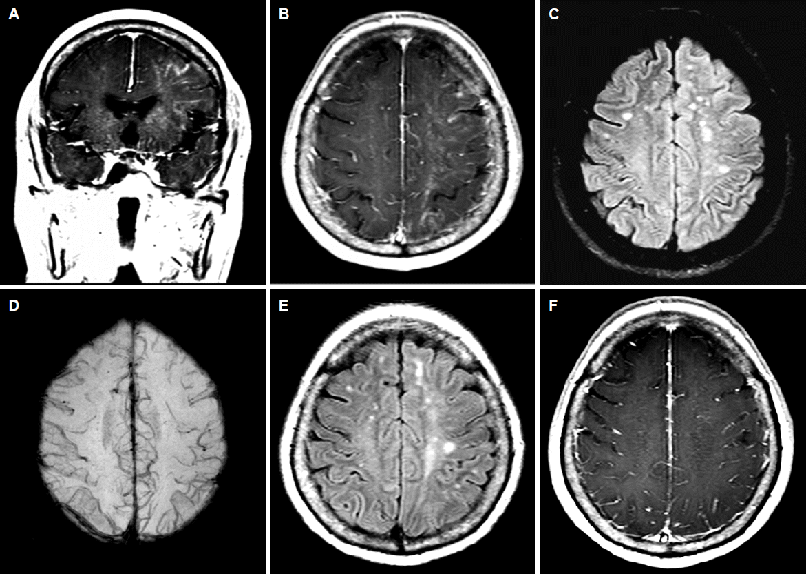
Magnetic resonance imaging (MRI) of the patient. Enhanced T1 weighted images showed an early parenchymal enhancement and diffuse sulcal enhancement in left cerebral hemisphere (A, B). T2 fluid-attenuated inversion recovery and susceptibility-weighted images showed nonspecific chronic ischemic changes without hemorrhagic lesions (C, D). Follow-up MRI after a week showed resolution of blood-brain barrier disruption of left hemisphere in contrast to previous study (E,F).
다양한 원인으로 발생하는 BBB 파괴는 심각한 뇌손상을 의미한다[1]. 외상뇌손상의 경우, 미세혈관손상 후 BBB 유지(integrity)에 관여하는 조절단백질의 감소에 따른 투과성변화에 의하며, 이는 출혈병변으로의 진행과 병변과 연관된 대뇌피질 증상악화의 예측인자이다[1]. 그러나, 본 증례처럼 일시적인 BBB 파괴를 보이나 좋은 예후를 보이는 경우도 있어[2] 예후 판단에 주의가 필요하다.
Acknowledgements
This study was supported by the Wonkwang University Research Grant in 2014.