눈깜박반사검사 이상소견으로 국소화된 우측 소뇌교뇌각종양

Right Cerebellopontine Angle Tumor Localized by Blink Reflex Abnormality

Article information

J Korean Neurol Assoc. 2016;34(1):86-87
Publication date (electronic) : February 1, 2016
doi : http://dx.doi.org/10.17340/jkna.2016.1.20
aDepartment of Neurology, Pusan National University Yangsan Hospital, Pusan National University Yangsan Hospital, Yangsan, Korea
bDepartment of Neurosurgery, Pusan National University Yangsan Hospital, Pusan National University Yangsan Hospital, Yangsan, Korea
cRisearch Institute for Convergence of Biomedical Science and Technology, Pusan National University Yangsan Hospital, Yangsan, Korea
채송화a, 신진홍a,c, 성순기b,c, 김대성,a,c
a양산부산대학교병원 신경과
b양산부산대학교병원 신경외과
c양산부산대학교병원 의생명융합연구소
Address for correspondence: Dae Seong Kim, MD  Department of Neurology, Pusan National University Yangsan Hospital, 20 Geumo-ro, Mulgeum-eup, Yangsan 50612, Korea  Tel: +82-55-360-2450 Fax: +82-55-360-2152 E-mail: dskim@pusan.ac.kr
received : July 27, 2015 , rev-recd : September 21, 2015 , accepted : September 21, 2015 .

64세 여자가 우측 삼차신경통으로 왔다. 병력청취와 신경학적진찰에서 얼굴감각이나 운동에 이상은 관찰되지 않았으나 우측 청력소실이 확인되었다. 눈깜박반사검사에서 우측 눈확위신경 자극시 양측 지연반사운동전위(R2)반응이 소실되어 있었고, 좌측 눈확위신경을 자극했을 때 반대측 R2반응이 소실되어 우측 가쪽연수병변을 시사하였다(Fig. 1). T1조영증강 뇌자기공명영상에서 우측 소뇌교뇌각종양이 뇌간을 압박하고 있었으며, T2영상에서 고신호강도가 가쪽연수의 상방까지 확장되어 있었다(Fig. 2). 수술소견에서 종양은 청신경과 얼굴신경을 침범하고 내이도, 소뇌, 삼차신경과 연접해 있었다. 수술 후 우측삼차신경통은 완전히 소실되었으나 이전에 없던 말초얼굴마비가 발생하였고 병리학적으로는 청신경초종(vestibular schwanoma)으로 확진되었다.
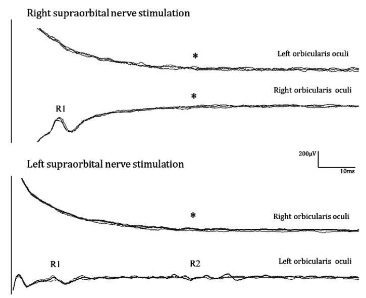
Figure 1.

Results of blink reflex test. Right supraorbital nerve stimulation provoked a normal ipsilateral R1 response, while neither ipsilateral nor contralateral R2 response (asterisks, upper panel) was observed. On left supraorbital nerve stimulation, normal ipsilateral R1 and R2 responses were observed, while contralateral R2 response (asterisk, lower panel) was not provoked. These findings strongly suggest a right lateral medullary lesion.

Figure 2.

Brainstem magnetic resonance imaging. (A) T1-weighted enhanced image shows well-marginated enhancing mass at right cerebellopontine angle (2.4×1.5×1.9 cm). (B) T2-weighted constructive interference in steady state image shows more detail in cochlear nerve area of brainstem by thin section (2 mm). Brainstem is compressed from the right side by the mass and accompanied by high signal intensity.

소뇌교뇌각종양에서 일반적으로 보이는 눈깜빡반사검사 소견의 경우 눈확위신경 자극시 양측 모두 반대측 R2의 변화와 함께 병변측 R1의 지연 또는 소실과 같은 변화도 같이 수반한다는 점으로 볼 때 [1,2], 본 증례에서 보였던 눈깜빡검사 이상소견은 일반적인 소뇌교뇌각종양에서 보이는 소견과는 차이가 있다. R2 반사경로는 동측 교뇌에서 연수 가쪽을 따라 하행한 후 양측의 얼굴신경핵으로 연결되므로 본 증례의 경우 우측 가쪽연수병변을 시사하였고, 이에 부합되는 소견을 영상과 수술을 통해 확인할 수 있었다.

References

1. Kimura J. The blink reflex as a clinical test. In : Michael J, ed. Electrodiagnosis in clinical neurology 5th ed. Vol. 17th ed. Philadelphia: Elsevier; 2005. p. 371–387.
2. Ongerboer de Visser BW, Kuypers HG. Late blink reflex changes in Lateral medullary lesions. Brain 1978;101:285–294.

Article information Continued

Figure 1.

Results of blink reflex test. Right supraorbital nerve stimulation provoked a normal ipsilateral R1 response, while neither ipsilateral nor contralateral R2 response (asterisks, upper panel) was observed. On left supraorbital nerve stimulation, normal ipsilateral R1 and R2 responses were observed, while contralateral R2 response (asterisk, lower panel) was not provoked. These findings strongly suggest a right lateral medullary lesion.

Figure 2.

Brainstem magnetic resonance imaging. (A) T1-weighted enhanced image shows well-marginated enhancing mass at right cerebellopontine angle (2.4×1.5×1.9 cm). (B) T2-weighted constructive interference in steady state image shows more detail in cochlear nerve area of brainstem by thin section (2 mm). Brainstem is compressed from the right side by the mass and accompanied by high signal intensity.