경추 뒤세로인대골화증 환자에서 컴퓨터단층촬영 및 자기공명영상
Cervical Spine CT and MRI Findings in a Patient with Ossification of the Posterior Longitudinal Ligament
Article information
61세 남자가 수년간의 경부통증 및 어깨관절 근력약화로 내원하였다. 생체징후는 정상이며 특이한 과거력은 없었다. 신경학적진찰에서 우측 어깨 굽힘 및 바깥회전시 근력저하(MRC grade 3)가 있었으나 감각이상 및 근위축은 없었다. 우측 어깨관절의 수동운동 제한은 없었다. 양측 상하지의 심부건 반사는 정상이었으며 Hoffmann징후는 보이지 않았다. 자기공명영상에서는 골화종괴를 뚜렷하게 확인할 수 없었으나 컴퓨터단층촬영에서 제 2경추에서부터 제 5경추까지 경추체 후방에 연속형의 경추후종인대의 골화가 보였다(Fig.). 이는 척추관협착 및 압박성 척수병증(compressive myelopathy)을 유발할 수 있다[1]. 저자들의 환자에서도 척수 압박 부위에 신호강도 증가부위가 보여 이는 척수 부종이 있음을 시사한다(Fig.) [2].
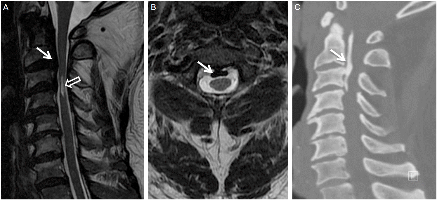
(A) The sagittal T2-weighted MRI shows bandlike low or no signal intensity of mass like lesion (white arrow) compressing spinal cord and short linear high signal intensity lesion (open arrow) posterior to C3-4 vertebral bodies. (B) The axial T2-weighted MRI shows focal rounded low or no signal intensity posterior to C2-3 vertebral bodies. (C) The sagittal reconstruction CT scan shows a long strip of ossification (white arrow) posterior to the C1-C3 vertebral bodies. White arrows in figure A and C indicate same level of vertebral bodies.