| J Korean Neurol Assoc > Volume 37(1); 2019 > Article |
|
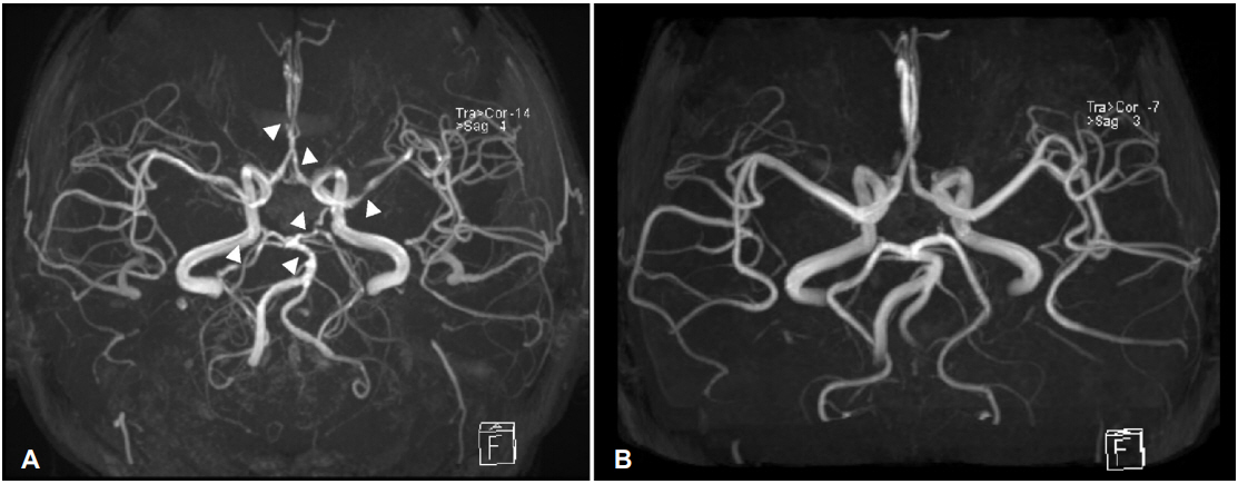
뇌들보(corpus callosum) 출혈은 외상, 색전성 뇌경색, 정맥혈전증, 혈관 기형 등에 의하여 유발되며 매우 드물다[1]. 27세 여성이 출산 일주일 후 갑자기 발생하여 1분 이내로 최고치에 이르는 두통을 주소로 내원하였다. 환자의 혈압은 174/103 mmHg이었으며, 뇌 컴퓨터단층촬영에서 뇌들보 출혈 소견이 관찰되었으나 뇌혈관은 정상이었다(Fig. 1-A). 하지만 입원 후 시행한 뇌혈관조영술에서 다발성 대뇌혈관협착 소견이 확인되었다(Fig. 1-B). 일주일 후 환자의 두통은 사라졌으나 뇌 자기공명혈관조영술에서 여전히 다발성 협착 소견이 관찰되었다(Fig. 2-A). 3개월 후 추적 뇌 자기공명혈관조영술에서 다발성 협착은 정상화되어 가역적뇌혈관수축증후군으로 진단하였다(Fig. 2-B) [2]. 이에 저자들은 가역적뇌혈관수축증후군에 의하여 유발되었으며 지연성 뇌혈관 변화를 보인 뇌들보 출혈에 대하여 보고한다.
Figure 1.
(A) Initial brain computed tomography (CT) scan showed corpus callosum hemorrhage (32×7×7 mm) with surrounding edema, however, brain CT angiography was normal. (B) The next day cerebral angiography was performed, which revealed diffuse multifocal arterial narrowing (arrows).

Figure 2.
(A) Brain magnetic resonance angiography (MRA) in a week disclosed multifocal stenosis of both anterior cerebral arteries (right A2, left A1), left middle cerebral artery (M1), both posterior cerebral arteries (right P2, left P1), and basilar artery (white triangles). (B) Repeated MRA in 3 months showed complete disappearance of the multifocal narrowing of the cerebral arteries.

- TOOLS
-
METRICS

-
- 0 Crossref
- 0 Scopus
- 3,777 View
- 70 Download
- Related articles
-
Reversible Cerebral Vasoconstriction Syndrome Induced by Blood Transfusion2020 November;38(4)
Reversible Cerebral Vasoconstriction Syndrome Associated with Severe Anemia2018 August;36(3)
Reversible Cerebral Vasoconstriction Syndrome after Heart Transplantation2017 November;35(4)
Steroid-Responsive Reversible Cerebral Vasoconstriction Syndrome2016 February;34(1)


 PDF Links
 PubReader
 ePub Link
 Full text via DOI
 Download Citation
 Print
Print


