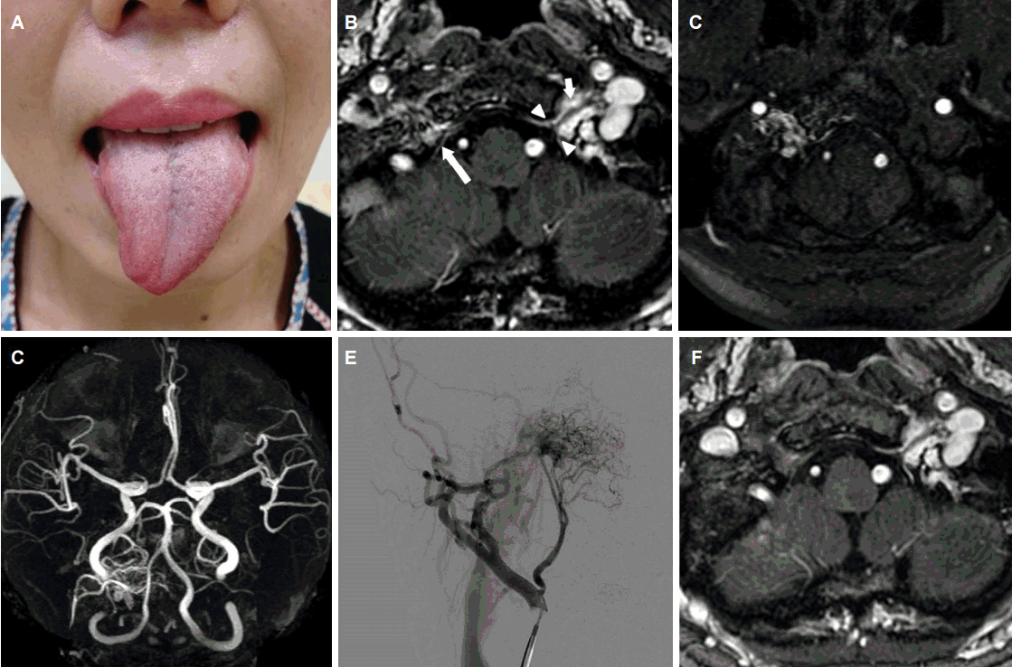
 | Figure.Photograph and imaging studies of the patient. (A) Tongue is mildly atrophied and deviated to the right during protrusion.(B) Fast spoiled gradient-echo (FSPGR) MRI demonstrates heterogenous hyperintense lesions around the right hypoglossal canal (long arrow). (B) Normal appearance of the left hypoglossal nerve (short arrow) and the borders of the hypoglossal canal (arrowheads) are depicted. (C) Source image of three-dimensional time-of-flight. (D) MR angiography shows high signal intensity around the right hypoglossal canal. MR angiography reveals abnormal vascularity lateral and superior to the right side of the foramen magnum. (E) Cerebral angiography demonstrates dural arteriovenous fistula supplied by the ascending pharyngeal artery with drainage into the internal jugular vein. (F) Follow-up MRI shows regression of the hyperintense lesions around the right hypoglossal canal. FSPGR; fast spoiled gradient-echo, MRI; magnetic resonance imaging, TOF; time-of-flight, MR; magnetic resonance. |
| J Korean Neurol Assoc > Volume 34(4); 2016 > Article |
|
52세 여성이 2주 전부터 구음장애가 있어 내원하였다. 두통이나 이명과 같은 외상은 없으나 4년 전 오른쪽 유방암 수술 후 완치된 병력이 있었다. 신체진찰에서 오른쪽 혀에 경미한 근위축이 있었고, 혀가 오른쪽으로 치우쳐 있었다(Fig. A). 근위축이 경미하고, 혀의 끝부분이 한 쪽으로 치우친 모습이 나타나 중추성 원인을 고려하였으나 뇌자기공명영상에서 뇌실질 이상은 보이지 않았고 우측 설하신경관 주위에 경계가 불분명한 조영증강이 나타났다(Fig. B). 자기공명혈관조영술에서 이상혈관을 시사하는 고신호 병변이 보였고(Fig. C, D), 뇌혈관조영술에서는 상행인두동맥에서 공급받고 내경정맥으로 연결되는 경막동정맥루가 설하신경관 부근에 있었다(Fig. E). 분할 정위방사선수술 후 설하신경마비는 회복되었고, 추적 영상에서 이상혈관을 시사하는 조영증강은 호전되었다(Fig. F). 단독설하신경마비는 종양이나 외상 또는 혈관이상에 의해 발생하는 경우가 흔하고, 설하신경관에 발생한 경막동정맥루가 원인인 경우는 드물다[1]. 경막동정맥루가 원인인 경우 색전술이나 방사선수술을 통해 치료가 가능하므로 두통이나 박동성이명과 같은 동반 증상이 없더라도 경막동정맥루를 확인하기 위해 적절한 검사가 필요하다[2].
- TOOLS
-
METRICS

-
- 0 Crossref
- 0 Scopus
- 6,675 View
- 117 Download
- Related articles
-
Congestive Myelopathy Due to Spinal Epidural Arteriovenous Fistula2019 November;37(4)
Delayed Isolated Hypoglossal Nerve Palsy after Submandibular Gland Surgery2016 May;34(2)


 PDF Links
 PubReader
 ePub Link
 Full text via DOI
 Download Citation
 Print
Print


