

77세 남자가 3일 전부터 발생한 전신 위약과 발열로 내원하였다. 기저 질환으로 범혈구감소증과 고혈압이 있었으며, 혈액 검사에서 혈소판 21,000/μL, 섬유소원(fibrinogen) 582 mg/dl, D-이합체(D-dimer) 3.70 mg/L, 프로트롬빈시간 31.0초로 ISTH criteria for disseminated intravascular coagulation 7점이 확인되었고, 폐렴막대균이 혈액 배양되어 정맥 내 세프트리악손을 투약하였다. 의식은 기면 상태였고 전신적으로 점상출혈이 확인되었다. 3년 전 시행한 뇌 자화율강조영상에서 미세출혈은 없었으나(Fig. A-C) 입원 11일째 시행한 뇌 컴퓨터단층촬영과 자기공명영상에서는 병변 주위 부종을 포함한 미세출혈이 소뇌, 뇌줄기 및 대뇌 전반적으로 분포해 있었다(Fig. D-I). 심각한 질병을 앓는 환자에서 다발성 뇌미세출혈이 발생할 수 있으며[1], 본 증례에서는 범혈구감소증 환자에서 중증 감염에 의한 파종성 혈관 내 응고 발생 이후 혈관 내 미세혈전과 소혈관 주변의 혈색소 침착으로 인해 광범위한 급성 뇌미세출혈이 발생한 것으로 추정된다[2].
| J Korean Neurol Assoc > Volume 41(3); 2023 > Article |

|
REFERENCES
1. Fanou EM, Coutinho JM, Shannon P, Kiehl TR, Levi MM, Wilcox ME, et al. Critical illness-associated cerebral microbleeds. Stroke 2017;48:1085-1087.


2. Kargiotis O, Safouris A, Magoufis G, Papageorgiou E, Fili M, Psychogios K, et al. Cerebral microbleeds: incidence, imaging characteristics, common and uncommon causes. J Neurosonol Neuroimag 2018;10:80-94.
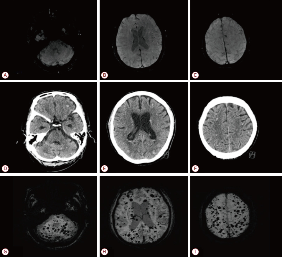
Figure.
MRI and CT images of the patient. SWI sequences showed no definite intracranial lesion on January 23, 2020 (A-C). Brain CT images (D-F), performed after 11 days of hospital day, December 14, 2022 showed multifocal lesions with high attenuation in the brain. And SWI sequences performed on the same day showed newly developed multiple scattered dark signal intensity lesions in bihemispheric cerebrum, cerebellum and ventricles (G-I). MRI; magnetic resonance imaging, CT; computed tomography, SWI; susceptibility-weighted-imaging.
- TOOLS
 PDF Links
 PubReader
 ePub Link
 Full text via DOI
 Download Citation
 Print
Print


-
METRICS

-
- 0 Crossref
- 0 Scopus
- 96 View
- 3 Download
-

- Related articles
-
Subacute Combined Degeneration in a Patient with Long-Term Oral Contraceptive Use2018 August;36(3)
The Association Between Hypertension and Cerebral Microbleeds in Patients With CADASIL2014 ;32(2)
Interictal Gastric Motility in Patients with Migraine2011 ;29(4)
Newly Developed Interest at Drawing in a Patient with Frontotemporal Dementia2011 ;29(1)
- Editorial Office
-
(ZIP 03163) #1111, Daeil Bldg, 12, Insadong-gil, Jongno-gu, Seoul, Korea
Tel: +82-2-737-6530 Fax: +82-2-737-6531 E-mail: jkna@neuro.or.kr
Copyright © 2024 by Korean Neurological Association.

