뇌 MRI에서 중간소뇌다리 신호 변화를 보이는 증례들
Differential Diagnosis of Middle Cerebellar Peduncle Signal Changes on Brain MRI
Article information
뇌 자기공명영상(magnetic resonance imaging, MRI)상 중간소뇌다리 신호 변화는 다양한 질환에서 관찰된다[1]. 액체감쇠역전회복영상에서 중간소뇌다리 신호 변화를 보이는 경우, MRI의 다른 영상이 질환의 감별에 도움이 된다. Fig. A는 갑자기 발생한 어지럼증과 휘청거림으로 내원한 환자의 뇌 MRI 영상이다. Fig. E 확산 강조영상의 양쪽 중간소뇌다리 신호변화와 겉보기확산계수지도를 통해 급성뇌경색으로 진단하였다. Fig. B는 진행하는 실조증과 자율신경이상 증세로 내원한 환자의 뇌 MRI로 Fig. F 액체감쇠역전회복영상에서 “십자가무늬빵”이 보였고, 다계통위축증으로 진단하였다. Fig. C는 갑작스러운 우측 하지마비로 내원한 환자의 증례로 Fig. G 확산강조영상에서 “지그재그테두리 징후”를 보였는데, 남동생의 확산강조영상에서도 같은 징후가 보였고, FMR1 음성이었다. 환자의 피부생검을 시행하여 신경세포핵내봉입체병(neuronal intranuclear inclusion disease)으로 진단하였다. Fig. D의 중간소뇌다리 신호변화는 만성뇌경색에 의한 축삭변성을 시사하는데[2], Fig. H T2강조영상에서 선명한 이전 상부뇌교뇌경색을 볼 수 있다. 위 4가지 다양한 질환 중 급성뇌경색 증례에서만 액체감쇠역전회복영상에서의 중간소뇌다리 신호 변화가 비대칭적이었다.
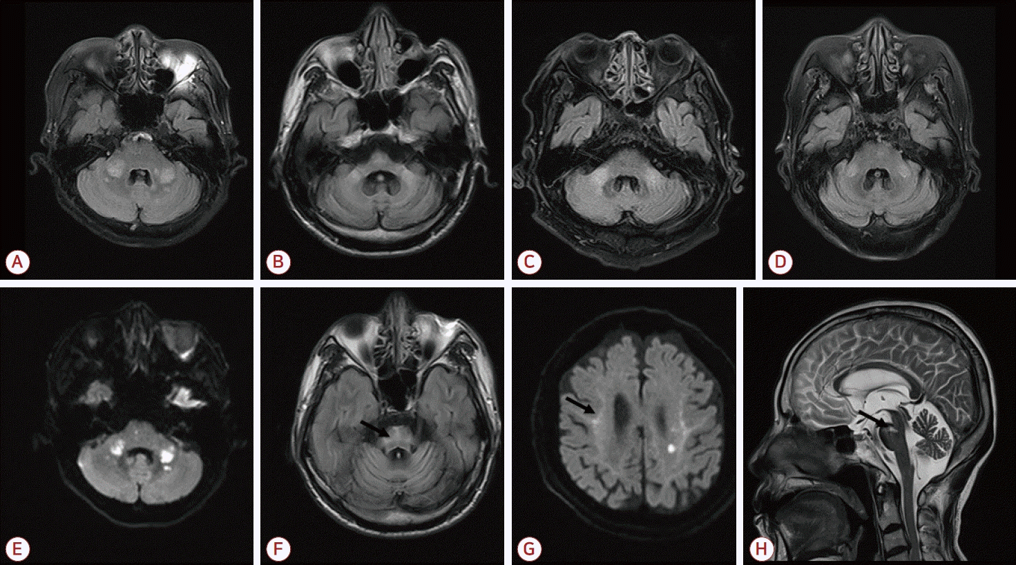
Upper row (A-D) shows bilateral high signal intensities on FLAIR axial images. E (diffusion-weighted axial image) disclosed asymmetric high signal intensities on middle cerebellar peduncles. Together with other brain images, we diagnosed the case acute posterior circulation infarction. F (FLAIR axial image) disclosed “hot cross bun” sign (arrow), which is suggestive of multiple system atrophy. G (diffusion-weighted axial image) revealed corticomedullary “zigzag edging” sign (arrow), which is suggestive of neuronal intranuclear inclusion disease. H (T2 sagittal image) showed a ventromedial upper pons infarction in chronic stage (arrow). FLAIR; fluid-attenuated inversion recovery.