혈관 내 치료로 개선된 다발경막동정맥루로 인한 급격한 신경학적 악화
Acute Neurologic Deterioration Associated with Multiple Dural Arteriovenous Fistula Improved by Endovascular Procedure
Article information
경막동정맥루(dural arteriovenous fistula, DAVF)는 후천적으로 발생하는 두개내 혈관기형 중 하나로 다양한 임상적인 경과와 증상을 보인다[1]. 뇌출혈은 DAVF의 가장 치명적인 합병증 중 하나이다. 따라서, DAVF의 중증도는 뇌출혈의 위험도를 반영하는 피질정맥 배출(cortical venous drainage)에 따라 분류하며 역행성 혈류로 인해 대뇌피질정맥의 울혈이 있는 경우 뇌출혈의 위험이 높다[1]. 하지만 뇌출혈이 없이 대뇌의 전반적인 피질정맥울혈만으로도 심각한 신경학적 이상을 보일 수 있으며, 적절한 치료로 신경학적 장애를 줄일 수 있다. 피질정맥울혈이 동반된 다발DAVF를 발견하고 적절히 치료한 증례를 경험하여 이를 보고하고자 한다.
증 례
74세 여자가 1개월 전부터 서서히 진행하는 인지저하로 응급실에 왔다. 2주 전부터는 구역감을 동반한 두통이 발생하였다. 간이정신상태검사는 19점이었고 무시증후군(neglect syndrome)과 함께 좌우혼동, 손가락인식불능, 연산능력저하(부분 거스트만증후군)도 동반되었다.
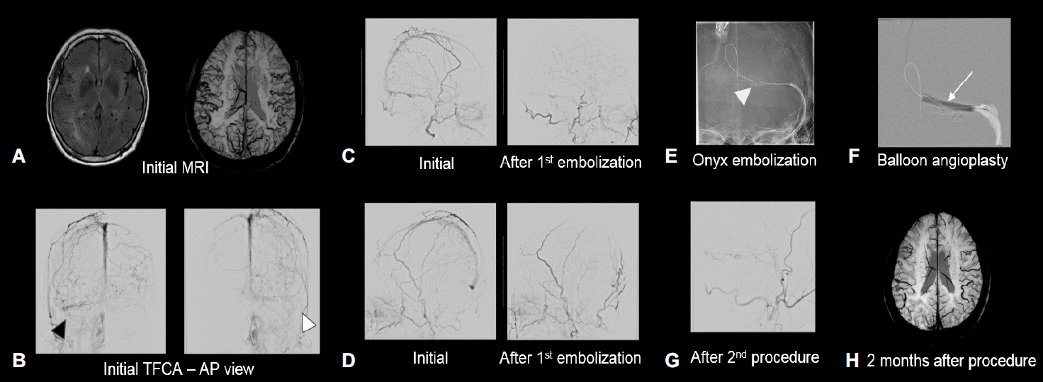
뇌 magnetic resonance imaging (MRI)에서 대뇌 전반에 걸친 뇌부종과 함께 대뇌 피질정맥울혈이 확인되었다(Fig. A). 입원 2일 뒤 급격한 의식 저하로 시행한 검사에서 우측 횡정맥동(transverse sinus)과 구불정맥동(sigmoid sinus)에 정맥혈전증이 확인되었다. 대퇴동맥경유뇌혈관조영(transfemoral cerebral angiography)에서 양측 표재측두동맥(superficial temporal artery, STA)와 후두동맥(occipital artery, OA), 중경막동맥(middle meningeal artery, MMA)에서 시작하는 다발DAVF와 양측 횡정맥동의 폐색이 확인되었다(Fig. B). 피질정맥울혈이 동반되어 있어 3형 Borden으로 분류할 수 있었다.

Brain magnetic resonance imaging and transfemoral cerebral angiography image. (A) FLAIR image showed diffuse brain vasogenic edema and SWI image showed venous engorgement in cortical vein (initial MRI). (B) Bilateral external carotid angiography showed multiple DAVF with venous congestion and occlusion of right transverse sinus (black arrowhead) and left transverse sinus (white arrowhead) (initial TFCA). (C) Angiography from right ECA and (D) left ECA, left image is initial angiography and right image is post embolization angiography using particle embolization. (E) Onyx embolization was done for torcular DAVF (white arrowhead) via left occipital artery approach. (F) An Aviator 4×30 mm balloon was used for dilatation in the left transverse sinus (white arrow). (G) Final angiography of the right ECA at the end of procedure showed improved venous drainage. (H) MRI was done 2 months after procedure and stent insertion in the left transverse sinus. Marked improvement in cortical venous engorgement was found. FLAIR; fluid attenuated inversion recovery, SWI; susceptibility weighted imaging, MRI; magnetic resonance imaging, DAVF; dural arteriovenous fistula, TFCA; transfemoral cerebral angiography, ECA; external carotid artery.
우측 외경동맥에서 시행한 조영술에서 피질정맥울혈이 좌측과 비교해 더 광범위하였고, 우측 뇌반구 증상이 더 우세하여 우측 외경동맥부터 색전술을 진행하였다. 피질정맥울혈을 유발하는 주요 혈류는 양측 OA, 우측 STA, 좌측 MMA에서 시작하였고 모든 DAVF에 대해서 색전술을 진행하는 것은 어려워 우측 OA, 우측 STA, 좌측 MMA, 좌측 OA 순서로 250-350 μm, 355-500 μm 크기 Contour 입자를 사용해 고식적색전술을 진행하였다(Fig. C, D). 색전술 이후 신경학적 호전이 확인되지 않았고 추적 영상에서 여전히 심한 피질정맥의 울혈이 확인되었다. 이에 왼쪽 OA로 접근하여 Onyx를 사용해 추가적인 색전술과 함께 정맥 배출의 빠른 개선을 위해 좌측 횡정맥동에 스텐트 삽입술을 동시에 진행하였다(Fig. E, F). 두 번째 시술 이후로 피질정맥울혈의 호전과 환자 의식이 호전되는 것이 확인되었다(Fig. G). 시술 시행 2개월 후 촬영한 MRI에서 피질정맥울혈의 감소가 확인되었다(Fig. H).
고 찰
다발DAVF는 70%에서 광범위한 뇌정맥혈전증이 보고되며 DAVF의 40%에서 뇌정맥혈전증이 동반되는 것과 비교해 더 높은 수치이다[2,3]. 다발DAVF의 발병과정에 대해서 여러 가지 가설이 있으나 가장 중요한 요인으로 뇌정맥혈전증이 제시된다[2]. 뇌정맥혈전증 이후에 재관류 과정에서 DAVF가 발생하게 되며 DAVF로 인해 변화된 혈류역학과 정맥고혈압은 다른 부위의 뇌정맥혈전증을 유발하며 이로 인해 또 다른 DAVF가 생성되어 다발DAVF로 진행한다. 이와 함께 항진된 혈관생성인자가 다발DAVF의 생성의 원인으로 제시되지만 직접적인 유발요인보다는 다발DAVF의 형성을 촉진하는 부가적인 요인으로 판단된다[2]. 다발DAVF에서 뇌정맥혈전증이 동반되는 빈도와 발병과정을 고려할 때 선후관계는 정확하지 않으나 다발DAVF과 뇌정맥혈전증은 밀접한 연관이 있음을 유추할 수 있다.
DAVF에서 뇌출혈 없이도 심한 신경학적 증상이 동반될 수 있으며 대부분 정맥울혈로 인해 유발된다고 알려져 있다[4]. 정맥울혈성뇌병증(venous congestive encephalopathy)으로 지칭하며 뇌 영상에서도 뇌의 구조적인 변화가 확인된다[4]. 정맥울혈의 정도는 정맥 역류를 통한 피질정맥울혈을 통해서 간접적으로 판단할 수 있으며 피질정맥울혈은 DAVF의 예후와 관련이 있는 것으로 알려져 있다[4]. 피질정맥울혈로 인해 구불구불하고 확장된 정맥이 보이는 것을 거짓정맥염(pseudophlebitis) 소견이라고 하며 거짓정맥염 소견의 정도에 따라 신경학적 증상과 심각도 그리고 예후와 연관이 있었다[4]. DAVF에서 피질정맥울혈의 유무와 뇌정맥협착은 밀접한 연관이 있는 것으로 생각된다. DAVF의 분류에 따라 뇌정맥협착이 동반된 빈도를 조사한 연구에서 1형 Cognard와 비교해 2형 Cognard와 피질정맥울혈이 동반된 DAVF에서 뇌정맥협착의 빈도가 높았다[5]. 피질정맥울혈이 없었던 DAVF에서 뇌정맥협착이 발생한 이후 정맥울혈이 발생하였고 급격하게 신경학적 증상이 악화되었던 보고도 확인되었다[1,5].
정맥울혈성뇌병증으로 인한 신경학적 증상이 중요한 이유는 가역적이기 때문이다. 1년 이상 진행하는 보행장애와 인지저하를 보인 환자에서 DAVF와 광범위한 피질정맥울혈이 확인되었고 색전술 이후에 호전이 확인된 증례 보고가 있다[6]. DAVF의 근본적인 치료는 모든 DAVF를 제거해주는 것이며 다발DAVF에서는 정맥 고혈압과 피질정맥울혈을 유발하는 DAVF를 색전술로 제거하는 것이 일차 치료이다. 하지만 조치가 필요한 모든 DAVF를 색전술로 제거하는 것은 어려우며 제한적인 색전술로는 충분한 치료효과를 보이지 않을 수 있다. 이러한 경우 다발DAVF와 뇌정맥협착의 선후 관계는 정확하지 않으나, DAVF에서 정맥울혈과 뇌정맥협착과의 연관성을 고려할 때 증상을 조절하기 위해 뇌정맥협착을 치료하여 정상적인 혈류의 회복을 도모하는 것이 도움이 될 수 있다[3,5,7].
다발DAVF 환자에게서 피질정맥울혈과 함께 신경학적 증상을 보일 때 적절한 색전술과 혈관내 시술을 통해 증상의 호전을 기대할 수 있어 빠른 진단과 시기 적절한 치료가 중요하겠다.