흉추경막동정맥루와 압박척수병의 동시 발현을 보인 자기공명영상
Magnetic Resonance Imaging in Co-Occurrence of Thoracic Spinal Dural Arteriovenous Fistula and Compressive Myelopathy
Article information
46세 남자가 2개월 전부터 시작한 양하지 위약으로 왔다. 위약은 수십 초간 간헐적 반복되다가 내원 1주일 전부터는 지속되었다. 배꼽 아래로 온도 및 진동감각저하를 확인하였다. 양하지의 근력 저하(Medical Research Council grade IV)와 발목간대경련이 있었다. 자기공명영상검사에서 흉추 4번에서 6번 수준에 경막동정맥루(Fig. A-C) 및 흉추 10번 부위의 압박척수병(Fig. A, B, D)이 있었다. 경막동정맥루에 대한 색전술과 감압척추고리판절제술을 병행하였고 3개월 뒤 근력은 완전히 호전되었다.
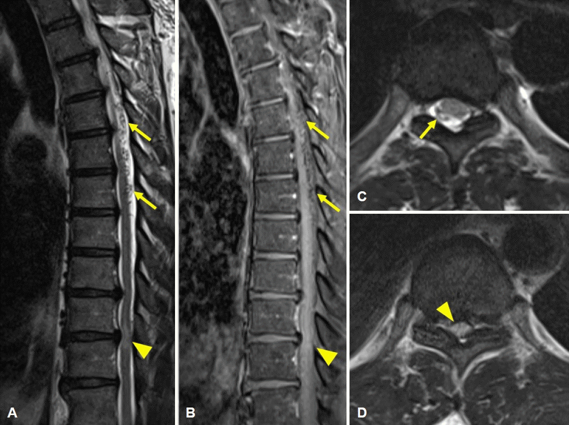
Engorged venous plexus with serpiginous intradural extramedullary flow voids were observed at T4-T6 level of spinal cord on T2 image (A, C, arrows) and T1 contrast enhanced image (B, arrows). Central canal stenosis due to disc protrusion and compressive myelopathy were observed at T9-10 level on T2 image (A, D, arrowhead) and T1 contrast enhanced image (B, arrowhead).
흉추의 압박척수병은 드물어 경추압박척수병에 비해 발병률이 1/10 수준이라고 보고된 바 있다[1]. 척수경막동정맥루는 척수의 혈관 기형 중 가장 흔한 형태로 흉추 6번에서 요추 2번 사이가 가장 흔한 병소로 추정된다[2]. 특히 간헐적 위약에 대한 평가에서 경막동정맥루의 가능성이 간과될 가능성이 있으므로 주의를 요한다.