뇌 아밀로이드 혈관병증-관련염증 증례를 통해 분석한 뇌척수액 아밀로이드 베타 및 플로르베타벤 뇌 양전자단층촬영 소견
Patterns of Cerebrospinal Fluid Biomarkers and Amyloid Positron Emission Tomography in a Patient with Cerebral Amyloid Angiopathy-Related Inflammation
Article information
Trans Abstract
Cerebral amyloid angiopathy-related inflammation (CAA-RI) is a distinct subset of cerebral amyloid angiopathy characterized by the auto-inflammatory response to amyloid-laden small arteries of cerebral cortex and leptomeninges. Clinical features include cognitive-behavioral change, headache, focal neurologic deficits and seizure. Because anti-inflammatory treatments can rapidly relieve neurologic symptoms, early diagnosis is critical. Herein, we report a CAA-RI case with distinct laboratory findings of a decreased cerebrospinal fluid amyloid beta 1-42 level and relatively reduced florbetaben uptake in the focal inflammatory lesion during the acute phase of CAA-RI.
뇌 아밀로이드 혈관병증-관련염증(cerebral amyloid angiopathy-related inflammation, CAA-RI)은 뇌 아밀로이드 혈관병증에서 보이는 미세출혈과 함께 백질의 국소 염증 및 부종이 동반되는 질병이다. 조직병리적으로 확진을 하지만, 조기에 정확하게 환자를 진단하기 위해 임상 증상과 뇌 자기공명영상검사 소견에 기반한 진단기준들이 제시되었고[1], 뇌척수액검사나 양전자단층촬영(positron emission tomography, PET)이 보조적으로 사용되고 있다. 저자들은 추정 CAA-RI로 진단된 환자의 급성기 뇌척수액에서 아밀로이드 베타-42 (amyloid beta 1-42, Aβ42) 및 타우(Tau)단백질 수치를 확인하였고, 불소화 포도당 뇌 양전자단층촬영(F-18 fluorodeoxyglucose PET, FDG-PET) 및 플로르베타벤 뇌 양전자단층촬영(F-18 florbetaben PET, FBB-PET)을 시행하였으며, 기존 보고와 비교를 통해 이들의 임상적 의미를 분석하였다.
증 례
70세 남자가 갑자기 발생한 혼동 증상을 주소로 응급실에 내원하였다. 환자에게 통풍 이외 다른 기저질환은 없었다. 약 3주 전부터 경미한 기억장애가 간헐적으로 있었다고 하며, 하의를 벗은 상태로 이웃집 벨을 누르는 이상행동을 보여 신고되었다. 응급실 도착 시 의식 수준은 명료하였으나 심한 혼돈을 보이고 있었으며, 가족들도 알아보지 못하고 질문에 적절한 대답을 하지 못하였다. 신경학적 검사에서 구음장애 및 언어장애가 확인되었다. 전반적인 언어 유창성이 감소되어 있었고, 따라 말하기, 이름대기장애가 두드러졌으며 음소 착어증이 빈번하게 관찰되었다. 그 외 뇌신경검사, 운동검사, 감각검사, 수막자극 징후는 정상이었으며 이상반사는 관찰되지 않았다.
경사에코영상에서 우측 측두-두정-후두엽 부위에 다발성 미세출혈(Fig. 1-A)과 우측 두정엽의 표재성 철 침착이 관찰되었고, fluid attenuated inversion recovery (FLAIR) 영상에서는 우측 내측 측두엽 피질하 백질 고신호 병변(Fig. 1-B)이 확인되었다. 우측 측두두정엽의 부종 및 국소 염증 소견이 각각 확산강조영상에서 고신호(Fig. 1-C), T1강조영상에서 뇌이랑을 따라 조영증강된 형태로 나타났다(Fig. 1-D). 현성확산계수 영상에서는 신호 변화가 뚜렷하지 않았다. 해당 부위는 FDG-PET검사에서 대사가 증가된 것으로 확인되었다(Fig. 1-E). 뇌파검사에서 우측 대뇌 반구에서 경련파(Fig. 2)가 지속적으로 관찰되어 비발작성 간질지속증이 진단되었으며, 레비티라세탐 및 발프로산을 정맥 주입하였다. 이후 혼돈 증상은 호전되었으나 언어장애는 지속되었다. 한글판 웨스턴 실어증 평가(Korean Western Aphasia Battery)검사 결과 중등도의 전도성 언어장애(aphasia quotient score, 56.8) 소견이 있었으나, 언어 실행증이 결과에 영향을 주었을 가능성이 있었다. 환자는 오른손잡이로 교차성 실어증(crossed aphasia) 가능성이 있을 것으로 판단되었다.
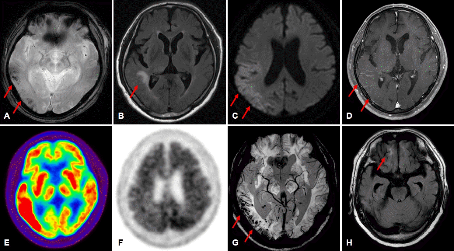
Findings of brain MRI, FDG PET and FBB PET. (A-F) At the first admission, there were multifocal microbleeds in the right temporo-parieto-occipital lobe (A, GRE sequence) and asymmetric white matter hyperintensity in the right medial temporal lobe (B, FLAIR sequence), as indicated by arrows respectively. Vasogenic edema in the right parieto-temporal area (arrows) was evident on the DWI image (C) with equivocal change on ADC map (not shown). There was gyral enhancement (arrows) in the right temporo-parietal cortex (D, Gd enhanced T1 weighted image) which implicates localized inflammation. Increased metabolism and relatively decreased amyloid uptake in the right parieto-temporal lobe were shown on the FDG-PET (E) and FBB-PET (F) images, respectively. (G-H) At the second admission, the extent of multifocal microbleeds (arrows) and superficial siderosis was increased in the right temporo-parieto-occipital area compared to the previous study (G, SWI sequence). There were diffuse brain atrophy slightly more prominent on the right medial temporal lobe (not shown) and increased hyperintensity lesion (arrows) in the right inferior frontal lobe (H, FLAIR sequence). MRI; magnetic resonance imaging, FDG; fluorodeoxyglucose, FBB; florbetaben, PET; positron emission tomography, GRE; gradient echo, FLAIR; fluid attenuated inversion recovery, DWI; diffusion weighted imaging, ADC; apparent diffusion coefficient, Gd; gadolinium, SWI; susceptibility weighted imaging.

Initial electroencephalography findings during the patient’s first admission. There were several episodes of electrographic seizures, which were 0.5 Hz lateralized periodic discharges in the right hemisphere evolving to 1-2 Hz sharpish lateralized periodic discharges.
FBB-PET검사에서는 대뇌피질 전반에 아밀로이드 침착이 확인되었다. 특히 좌측 후측 대상피질 및 두정피질에서 아밀로이드 침착이 두드러졌고, 국소 염증을 보였던 우측 측두-두정엽 부위에서는 상대적으로 감소되어 보였다(Fig. 1-F). 뇌척수액검사에서 세포 수 및 단백질 수치는 정상이었다. 뇌척수액 Aβ42는 감소되어 있었고(109.98 pg/mL) 총 Tau는 증가되어 있었다(974.27 pg/mL). 인산화 Tau (phosphoraylated serline 199; 1.16 pg/mL)는 정상 범위로 확인되었다. 아포지단백E 유전자형은 E3/E4로 확인되었다. 환자는 추정 CAA-RI로 진단되어 5일간 고용량 스테로이드(methylprednisolone 1 g, IV) 치료를 받았다. 이후 환자의 인지장애는 점차 호전을 보였고, 한국 간이정신상태검사로 평가하였을 때 치료 전 17점에서 치료 후 28점으로 향상되었다.
스테로이드는 경구로 복용하면서 서서히 감량하던 중이었으나, 퇴원 2개월 후 언어장애와 이상행동이 재발하였다. Susceptibility weighted imaging영상에서 우측 측두-두정-후두엽과 소뇌 반구에 다발성 미세출혈과 표재성 철 침착이 증가되어 보였고(Fig. 1-G), FLAIR영상에서 우측 아래 전두엽 영역으로 고신호 병변이 새롭게 관찰되었다(Fig. 1-H). 뇌파검사에서는 우측 두정-후두엽 부위에서 경련파가 확인되었다. 레비티라세탐, 발프로산, 라코산, 페람파넬 등 여러 항경련제를 투약하였음에도 뇌파 감시 상에서 국소 경련파가 지속되었다. 난치성 경련중첩상태로, 기관내삽관 하에 케타민 및 프로포폴 지속 주입을 시작하였고, 페노바비탈, 클로바잠, 토피라메이트를 추가하였다. 입원 14일 후 뇌파 소견이 호전되어 항경련제 및 진정제를 감량하였다. 다만 인지장애는 지속되었고 공격적인 행동이 관찰되었다. CAA-RI 재발로 판단되었고, 동반된 흡인성 폐렴이 호전된 후 고용량 스테로이드 요법을 시행하였다. 행동 조절을 위해 올란자핀을 추가하였고 레베티라세탐 및 페람파넬은 중단하였다. 이후 인지장애는 호전을 보였으며, 한국 간이정신 상태검사 점수가 15점까지 떨어졌던 것이 23점으로 향상되었다. 퇴원 이후에는 도네페질, 저용량 경구 스테로이드, 항경련제(발프로산, 페노바비탈)를 유지 중에 있다.
고 찰
본 증례는 급성 의식장애, 인지행동 변화, 언어장애, 비발작성 간질지속증이 발현된 환자에서 추정 CAA-RI가 진단된 예로, 고용량 스테로이드 치료 후 임상적 관해가 있었으나 2개월만에 재발한 매우 드문 사례이다. CAA와는 달리, CAA-RI는 가역적 뇌병증의 증상과 뇌 자기공명영상에서 확인되는 뇌 국소 염증 및 부종을 특징으로 하며, 대부분 고용량 스테로이드에 잘 반응하여 약 70%의 환자에서 임상 및 영상학적 호전을 보인다[2]. 일부 문헌에서는 추가로 면역억제제(예; 메토트렉세이트, 미코페놀레이트 모페틸, 사이클로포스파미드)를 투약하거나 면역글로불린을 투약하여 임상 호전을 보인 경우도 보고하였다[2]. 예후는 보고 마다 편차가 있으며, 뇌출혈 또는 기회감염으로 사망하는 경우가 있었고(14-33%) [1,3], 생존하는 경우 48%에서 무증상이거나 경미한 신경학적 후유증을 보였다[1]. 재발률은 3-43%까지 다양하게 보고되나1,3,4 스테로이드를 투약하였던 경우에는 치료하지 않았던 경우보다 재발률이 두 배 더 낮았다[3]. 본 증례에서는 첫 증상으로부터 고용량 스테로이드 투약까지 약 3주가 소요되었으며, 투약 후 2개월 만에 재발이 있었고, 두 번째 스테로이드 충격요법의 반복 후에 다시 임상 호전을 보였다.
현재의 임상적-영상학적 진단기준 이외의 새로운 진단적 가치를 갖는 생물학적 표지자를 확인하기 위해 저자들은 뇌척수액에서 Aβ42, 총 Tau, 인산화 Tau단백 수치를 확인하였고, 아포지단백E의 유전자형검사를 시행하였다. 추가로 환자의 급성기 뇌 병변에서 대사 상태 변화 및 아밀로이드 침착 정도를 양전자단층촬영검사를 통해 확인하였다. 상기 표지자들의 진단 가치에 대해 알아보기 위해 2013년도부터 2019년도까지 보고된 국외 논문 9개를 고찰하였다(Supplementary Table). 대부분의 연구에서는 뇌척수액 Aß의 감소, 총 Tau 및 인산화 Tau의 증가를 보이고 있었다. 다만 이러한 경향은 알츠하이머치매나 CAA에서도 비슷하게 보고되는데, 이들의 차이점을 분석한 논문에서는 CAA와 비교 시 뇌척수액의 Aβ42가 CAA-RI에서 더 유의미하게 낮았으며, 알츠하이머치매와 비교 시 Aβ40, 총 Tau 및 인산화 Tau가 CAA-RI에서 더 유의미하게 낮았다[5]. 또한 항아밀로이드항체를 이용한 과거 임상 연구들에서 보고된 부작용 중에 하나인 아밀로이드 관련 영상학적 비정상 소견(amyloid related imaging abnormalities)과 CAA-RI가 유사한 표현형을 보인다는 점에서 Aβ가 CAA-RI의 발생 기전에 중요한 역할을 할 것임을 유추할 수 있다. 이러한 가설은 몇 건의 보고들에 의해 더욱 지지를 받는데, 급성기 CAA-RI 환자에서 항Aβ42항체의 역가가 증가되어 있었으며(Supplementary Table), 임상적 관해기에 재측정한 경우 항체의 역가가 정상 범위로 감소된 것이 확인되었다[6,7]. 본 증례에서는 항Aβ42항체를 검사하지 못하였으나 환자의 급성기 뇌척수액 검체에서 Aβ42 수치가 현저하게 감소되어 있음을 확인하였다.
CAA-RI 환자에서 아포지단백E 대립유전자의 아형을 비교하였을 때 두 개의 E4 동종아형을 가지는 경우가 45.8%, 한 개 이상의 E4 아형을 가지는 경우가 62.5%로 E4 아형과의 연관성이 확인되었으며(Supplementary Table), 본 증례의 환자는 E3/E4 아형으로 확인되었다.
CAA-RI 환자에서 FDG-PET검사를 시행한 증례는 드물었는데, 본 증례에서는 질병의 급성기에 뇌 병변에서 대사가 증가된 소견이 있었으며, 다른 증례 보고에서는 두 명의 환자에서 각각 스테로이드 투약 후 또는 증상 호전 뒤에 검사를 시행하였을 때 뇌 병변 부위에서 대사가 감소된 것이 확인되었다[8]. 본 증례에서 급성기 CAA-RI 환자의 FBB-PET검사 소견에서는 뇌 국소 염증 부위에서 아밀로이드 침착이 상대적으로 감소되어 보였는데, 이는 피츠버그 화합물 B (C-11 Pittsburgh compound B, PiB) PET영상을 통해 분석한 두 건의 보고에서 급성기 뇌 염증 병변에서 아밀로이드 침착이 감소되었던 것과 일치한다[4,9]. 이러한 현상은 gantenerumab과 같은 항Aβ42항체의 치료효과를 분석한 보고에서도 비슷하게 관찰되었는데, 항체를 투약한 군에서는 PiB-PET검사에서 아밀로이드 침착이 감소됨이 확인되었다[10]. 즉 CAA-RI 환자의 국소 뇌 병변에서 관찰되는 아밀로이드 침착의 감소 형태는 Aβ에 작용하는 자가 염증반응에 의한 Aβ 제거현상(clearance)과 연관된 것으로 판단되며, CAA-RI의 관해기에 검사한 PiB-PET에서는 이러한 소견이 관찰되지 않았다[8].
저자들은 CAA-RI로 진단된 환자에서 급성기의 특징적인 소견으로 뇌척수액 Aβ42의 감소, FDG-PET에서 병변의 대사 증가, FBB-PET에서 병변의 아밀로이드 침착 감소가 있었음을 보고하며, magnetic resonance imaging 이외의 상기 검사들이 CAA-RI의 정확하고 빠른 진단과 치료계획 수립에 도움을 줄 수 있음을 강조하고자 한다.
Supplementary Material
Supplementary Table.
Review of reported cases of CAA-RI by comparing the results of CSF markers, ApoE genotypes, FDG-PET and amyloid PET studies