J Korean Neurol Assoc > Volume 39(1); 2021 > Article
종양의 직접 색전에 의한 급성 대뇌동맥 폐색

Abstract

In acute stroke, emboli are mostly composed of thrombi from artery, cardiac chamber, valve and vein. Non-thrombotic emboli are sometimes difficult to identify the origin. According to the increased number of cancer patients, now 10% of stroke patients have a cancer. However, the potential mechanisms of stroke in patients with cancer are various. We presented a case of serious acute arterial occlusion with a tumor embolus, which was revealed by histopathologic analysis of retrieved emboli during mechanical thrombectomy.

뇌졸중 환자 10명 중 1명은 암을 동반질환으로 가지고 있다[1,2]. 암 환자에서 뇌졸중은 응고병증, 비감염심내막염, 심부정맥혈전증과 같은 병인으로 흔히 설명된다[2,3]. 그러나 여전히 암 환자 뇌졸중의 48%는 원인불명(undetermined etiology)으로 보고되고[3], 이 중에서 종양 자체가 혈전 및 색전으로 작용하여 뇌졸중을 발생시키기도 하는데, 실제 조직학적으로 증명된 경우는 극히 드물다. 최근에 허혈뇌졸중 치료 기술의 발달로 직접 혈전 조직을 얻을 수 있게 되면서 뇌졸중의 원인을 밝히려는 노력이 계속해서 시도되고 있는데, 본 증례는 급성 뇌졸중 환자에서 동맥내혈전제거술을 이용하여 얻어진 조직검사를 통해, 종양 자체가 직접 혈관을 막을 수 있음을 보여주는 드문 증례를 보고하고자 한다.

증 례

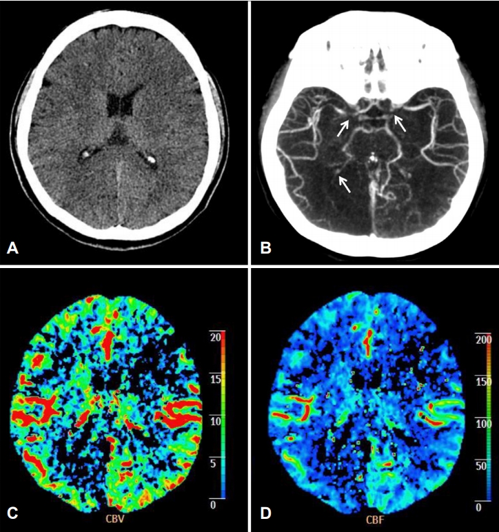
56세 여성 환자로 의식저하와 우측 편마비(Medical Research Council 등급 II)를 주소로 전원되었다. 평소 건강했던 자로 병원에 오기 3시간 전 집에서 쓰러져 있는 채 발견되었다. 신경계진찰에서 의식상태는 혼미, 좌측으로 안구편향 및 우측 편마비였고 미국 국립보건원 뇌졸중 척도에서 17점을 보였으며, 호흡이 불안정하여 기관을 삽관하였다. 뇌 computed tomography (CT)에서 좌측 전두엽의 저음영(Alberta Stroke Program Early CT Score 8), 우측 후두엽의 저음영을 보였다(Fig. 1-A). 뇌 CT혈관조영검사에서 양측내경동맥 원위부 폐색과 우측 후대뇌동맥 폐색이 관찰되었다(Fig.1-B). 저음영이 의심되는 부위로 뇌관류 결손을 확인할 수 있었다(Fig. 1-C, D). 증상 발생 3시간으로 정맥내조직 플라스미노젠활성제(intravenous tissue plasminogen activator) 투입 후 기계적 동맥내혈전제거술을 시행하였다.
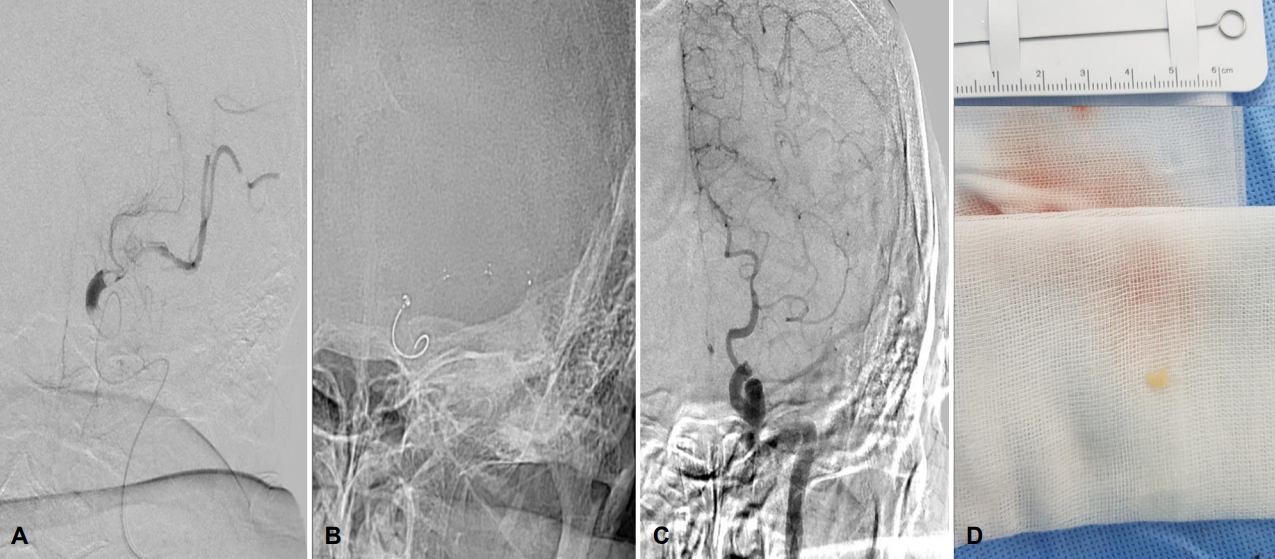
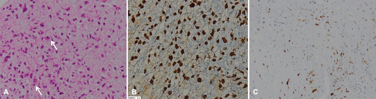
뇌혈관조영술에서 좌측 내경동맥 원위부 폐색을 확인하였고(Fig. 2-A) 좌측 내경동맥에서 Solitaire™ (Medtronic, Minneapolis, MN, USA) (4-20 mm) 2차례, Solitaire™ (Medtronic) (6-40 mm) 10차례 혈전제거술(Fig. 2-B)을 시행하였으나 재관류에 실패하였다(Fig. 2-C). 우측 내경동맥 원위부와 중대뇌동맥의 협착 소견이 보이나 혈류는 유지되어 100여 분간의 시술시간 및 환자 상태를 고려하여 시술을 종료하였고 시술 후 thrombolysis in cerebral infarction 등급 0이었다. 혈전제거술에서 일부 얻어진 혈전은 일반적인 색과 달리 백색의 조그만 크기(4 mm, Fig. 2-D)였고, 조직염색을 위해 포르말린 용액에 바로 고정을 하고 염색하였다. 환자는 시술 후에도 의식 회복하지 못하였으며, 2시간 후 추적 뇌 CT에서 양측 전두엽, 측두엽과 우측 후두엽의 허혈병변을 확인하였고, 급성 뇌부종으로 진행하여 당일 사망하였다. 혈전 조직에 대한 헤마톡실린과 에오신염색(Fig. 3-A) 결과는 비전형적인 간엽세포의 증식, 다형태성, 과염색성 등 악성종양의 특징을 보여 종양의 기원을 확인하기 위한 추가적인 면역염색과정으로 시토케라틴(상피세포, 음성), 에스-100(신경세포, 음성), 비멘틴(중간엽, 양성, Fig. 3-B)에 대해 시행하여 육종을 확인하였고 이 중에서도 액틴(평활근, 음성), 데스민(골격근과 횡문근, 양성, Fig. 3-C) 결과로 횡문근육종에 합당하였다.

고 찰

본 증례는 급성 뇌졸중 환자에서 동맥내혈전제거술을 통해 얻은 혈전을 조직염색검사를 시행하여 종양 자체가 혈전 및 색전으로 작용하였음을 확인하였다. 그동안 심장횡문근육종, 좌심방 연골육종 환자에서 급성 뇌경색이 발생한 보고가 있었으나 심장 초음파와 심혈관 컴퓨터단층촬영을 통해 종양의 존재만을 확인한 경우로 종양에 의한 2차적 혈전인지 혈전 자체가 종양인지에 대한 의문이 있어왔다[4-6]. 최근 혈전제거술이 일반화됨에 따라 종양 자체가 혈전 및 색전으로 직접 작용하여 급성 뇌졸중 일으킨 미분화육종, 편평세포암종 등 드물게 보고되고 있다[7-9]. 따라서 본 증례는 종양 색전에 의한 직접 혈관 폐색이 가능하다는 점을 시사한다.
육종은 약 30%에서는 심장기원으로 보고되고 있으나 전신 어디에서든 발생할 수 있다[8]. 본 증례 환자의 과거력에서 건강했다고 하였고, 흉부 X-선촬영, 응급 혈액검사 등에서는 이상 소견은 없었으며 심전도와 심장표지자에서 심방세동이나 심근경색과 같은 심장질환은 없었다. 환자 예후가 좋지 않아 심장기원의 유무 및 종양의 전이 여부는 확인할 수 없었다. 뇌혈관조영술에서도 양측 내경동맥 원위부의 상태를 고려하였을 때 기저질환으로 두개내 죽경화를 동반했을 가능성은 있으나 종양을 의심할 만한 소견은 보이지 않았다. 하지만 동맥내혈전제거술과 조직염색을 통해 암과 관련된 흔치 않은 병인을 확인할 수 있었고 뇌졸중의 병인을 밝혀내는 중요한 과정임을 말해준다[10].
암은 뇌졸중과 심방세동, 비만, 흡연과 같은 공통된 위험요인을 가지고 이와 관련된 응고병증과 항암 치료로 인해 뇌졸중의 위험성에도 영향을 주고 있으며 고령 등 암을 동반하는 뇌졸중 환자가 증가함에 따라 정확한 원인 규명이 필요하다. 따라서 혈전 색전증의 원인이 종양 자체임을 밝혀낸 증례는 중요한 의의가 있기에 동맥내혈전제거술과 함께 조직염색검사의 중요성 또한 강조하며 이에 대해 보고한다.

REFERENCES

1. Navi BB, Iadecola C. Ischemic stroke in cancer patients: a review of an underappreciated pathology. Ann Neurol 2018;83:873-883.
crossref pmid pmc
2. Bang OY, Chung JW, Lee MJ, Seo WK, Kim GM, Ahn MJ, et al. Cancer-related stroke: an emerging subtype of ischemic stroke with unique pathomechanisms. J Stroke 2020;22:1-10.
crossref pmid pmc
3. Schwarzbach CJ, Schaefer A, Ebert A, Held V, Bolonrese M, Kablau M, et al. Stroke and cancer: the importance of cancer-associated hypercoagulation as a possible stroke etiology. Stroke 2012;43:3029-3034.
pmid
4. Oizumi H, Tanaka R, Shimura H, Sasaki K, Koike H, Hattori N, et al. A case of cerebral embolism with metastatic chondrosarcoma in the left atrium. J Stroke Cerebrovasc Dis 2011;20:79-81.
crossref
5. Ashraf T, Day TG, Marek J, Hughes M, Giardini A. A triad: cardiac rhabdomyosarcoma, stroke and tamponade. Pediatr Cardiol 2013;34:771-773.
crossref pmid
6. Caballero PE. Left atrial sarcoma presenting as cerebral infarction. Neurologist 2008;14:131-133.
crossref pmid
7. Lemus HN, Lu C, Shoirah H, Shigematsu T, Liang J, Van denakker T, et al. Direct tumor embolism presenting as an acute ischemic stroke. Neurol Clin Pract 2019;9:490-493.
crossref
8. Byon JH, Kwak HS, Chung GH, Jang KY. Acute stroke from tumor embolus in a patient with cardiac sarcoma: aspiration thrombectomy with penumbra catheter. Interv Neuroradiol 2016;22:88-90.
crossref pmid pmc
9. Fukami Y, Yamaguchi K, Miyasaki A, Negoro M. Acute ischemic stroke due to undifferentiated sarcoma: a case report and literature review. Intern Med 2019;58:115-118.
crossref pmid
10. Ahn SH, Hong R, Choo IS, Heo JH, Nam HS, Kang HG, et al. Histologic features of acute thrombi retrieved from stroke patients during mechanical reperfusion therapy. Int J Stroke 2016;11:1036-1044.
crossref pmid

Figure 1.
Brain computed tomography angiography and perfusion computed tomography. (A) Initial non contrast brain computed tomography shows left frontal lobe and right occipital lobe low density. (B) Brain computed tomography angiography (arrows) reveal the occlusion of bilateral distal internal carotid artery and right posterior cerebral artery. (C) Cerebral blood volume and (D) cerebral blood flow of perfusion computed tomography shows decreased in left frontal and right occipital lobes.
jkna-39-1-30f1.jpg
Figure 2.
Digital subtraction angiography and specimen of retrieved embolus. (A) First digital subtraction angiography shows left distal internal carotid artery (ICA) occlusion. (B) Although 12 times trials of stent retrieval procedure, (C) distal ICA was not recanalized. (D) A white-colored specimen with size of 4 mm is retrieved.
jkna-39-1-30f2.jpg
Figure 3.
Histopathologic findings of retrieved embolus. (A) Hematoxylin-eosin staining (×400) shows characteristic such as pleomorphism, hyperchromatism, and frequent mitoses (arrows) that indicate a cancer. It consists of loose, myxoid soft tissue and each cell was round to oval, spindle, polygonal and stellate shape. These finding suggests a malignant tumor of non-epithelial origin. (B) By strong immune reactivity for positive vimentin (×400) and (C) positive desmin staining (×100), rhabdomyosarcoma is confirmed finally.
jkna-39-1-30f3.jpg


ABOUT
BROWSE ARTICLES
EDITORIAL POLICY
FOR CONTRIBUTORS
Editorial Office
(ZIP 03163) #1111, Daeil Bldg, 12, Insadong-gil, Jongno-gu, Seoul, Korea
Tel: +82-2-737-6530    Fax: +82-2-737-6531    E-mail: jkna@neuro.or.kr                

Copyright © 2024 by Korean Neurological Association.

Developed in M2PI

Close layer