

62세 남자가 갑자기 발생한 의식저하로 내원하였다. 신경계진찰에서 빛반사는 정상이었으나 의식 수준은 혼미하여 글래스고혼수 척도는 7점이었다. 혈압약만을 복용 중이었고 활력징후와 혈액검사는 정상이었다. 뇌 자기공명영상 확산강조영상에서 양측 정중곁 시상과 중뇌에 고신호이고 겉보기확산계수지도에서 저신호인 병변이 보였고, 가톨리늄 조영제를 사용하여 촬영한 혈관조영영상에서 Percheron 동맥 폐색 의심 소견이 보였다(Fig.). 뇌경색으로 진단하여 항혈전제 복용을 시작하였고 2일 후 의식은 호전되었으나 양안의 하방주시장애와 기억력저하가 확인되었다.
| J Korean Neurol Assoc > Volume 38(4); 2020 > Article |

|
REFERENCES
1. Arauz A, Patiño-Rodríguez HM, Vargas-González JC, ArguellesMorales N, Silos H, Ruiz-Franco A. Clinical spectrum of artery of percheron infarct: clinical–radiological correlations. J Stroke Cerebrovasc Dis 2014;23:1083-1088.


2. Lazzaro NA, Wright B, Castillo M, Fischbein NJ, Glastonbury CM, Hildenbrand PG, et al. Artery of percheron infarction: imaging patterns and clinical spectrum. AJNR Am J Neuroradiol 2010;31:1283-1289.
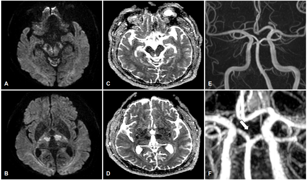
Figure.
(A, B) MRI DWI shows high signal intensity lesions in the midbrain and bilateral paramedian thalamus with low signal intensity in (C, D) ADC images. (E) Conventional MRA shows no significant stenosis or occlusion on basilar and posterior cerebral artery. (F, arrow) Gadolinium contrast-enhanced MRA shows suspicious occluded artery of Percheron. MRI; magnetic resonance imaging, DWI; diffusion weighted images, ADC; apparent diffusion coefficient, MRA; magnetic resonance angiography.
- TOOLS
 PDF Links
 PubReader
 ePub Link
 Full text via DOI
 Download Citation
 Print
Print


-
METRICS

-
- 0 Crossref
- 0 Scopus
- 2,785 View
- 64 Download
- Related articles
- Editorial Office
-
(ZIP 03163) #1111, Daeil Bldg, 12, Insadong-gil, Jongno-gu, Seoul, Korea
Tel: +82-2-737-6530 Fax: +82-2-737-6531 E-mail: jkna@neuro.or.kr
Copyright © 2026 by Korean Neurological Association.

