특발두개내압상승의 Magnetic Resonance Imaging 징후
Magnetic Resonance Imaging Signs, as a Dandy Compass Pointing to Idiopathic Intracranial Hypertension
Article information
19세 여자가 시야흐림과 두통으로 안과를 방문하였다. 안저검사에서 양안 시신경유두부종이 있었고, 뇌 magnetic resonance imaging (MRI)에서 구조적 뇌병터와 수두증은 없었다. 고용량 스테로이드 치료 후 시야흐림은 호전되었으나 두통이 지속되어 신경과에 의뢰되었다. 신경계진찰에서 국소신경학적 결손은 없었고, 신체비만지수가 27.4였다(키 162 cm, 몸무게 72 kg). 초기 뇌 MRI를 재검토한 결과, 안구 후면의 편평화와 부분빈안장(partial empty sella)이 보였다(Fig.). 뇌척수액검사에서 정상 뇌척수액 구성과 두개내압 >300 mmH2O로 특발두개내압상승으로 진단하였다[1]. 특발두개내압상승의 영상 징후는 안구 후면의 편평화, 시신경집(optic nerve sheath) 팽창, 시신경 비틀림, 부분빈안장이 있다[1,2]. 개개의 징후는 특발두개내압상승 진단기준을 대체할 수 없고 민감도가 낮은 제한이 있지만, 진단 접근의 맥락에서 훌륭한 지침으로 활용할 수 있다.
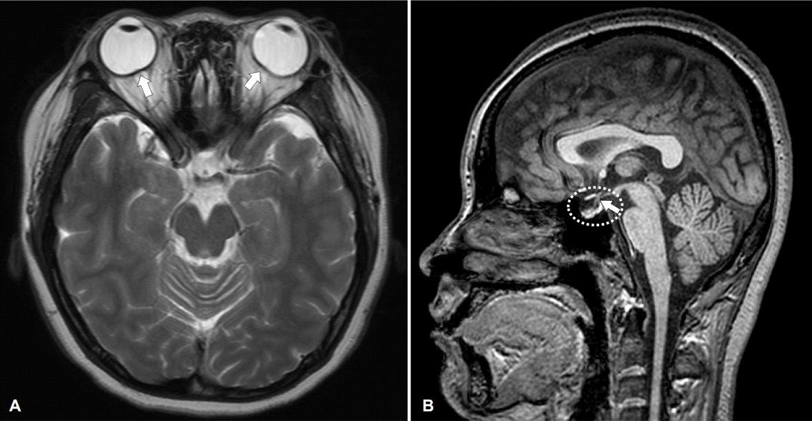
Magnetic resonance imaging (MRI) signs pointing to idiopathic intracranial hypertension. (A) Axial T2 weighted image shows posterior flattening of bilateral optic globes (arrows). (B) Sagittal T1 weighted image shows partially empty sella (dotted oval) with infundibulum sign (arrow).