

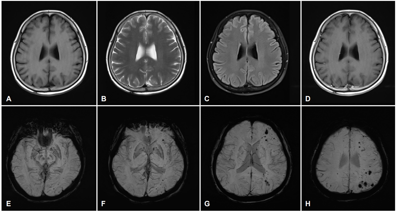
59세 여자가 어지러움, 두통과 안면통으로 병원에 왔다. 뇌 자기공명영상에서 T1강조영상, T2강조영상, 액체감쇠역전회복영상, 조영증강 T1강조영상에서 이상이 없었고(Fig. A-D), 감수성강조영상에서 다양한 크기의 다발성 저신호강도가 광범위하게 보였다(Fig. E-H). 자기공명영상으로 다발성 뇌해면기형으로 진단하였다. 가족성뇌해면기형의 진단기준은 5개 이상의 병변, 가족 중에 2명 이상에서 발현, 관련 유전자의 돌연변이 중에 한 가지만 만족하여도 진단할 수 있다[1]. 가족성뇌해면기형은 자기공명영상을 기준으로 4가지 형으로 분류한다[2]. 제4형은 점 모양의 저신호 병변이 감수성강조와 기울기에코영상에서 관찰되나, T1과 T2강조영상에서는 병변이 보이지 않는다[2]. 미세출혈의 크기가 다양하고, 고혈압이나 뇌아밀로이드혈관병증으로 인한 미세출혈 호발 부위 외의 병변이 있는 것은 뇌해면기형과 관련될 가능성이 높다. 본 증례는 다양한 크기의 많은 병소를 가진 다발성 뇌해면기형으로, 영상학적으로는 가족성 제4형으로 여겨진다.
| J Korean Neurol Assoc > Volume 37(3); 2019 > Article |

|
REFERENCES
1. Mespreuve M, Vanhoenacker F, Lemmerling M. Familial multiple cavernous malformation syndrome: MR features in this uncommon but silent threat. J Belg Soc Radiol 2016;100:51.


2. de Souza JM, Domingues RC, Cruz LC Jr, Domingues FS, Iasbeck T, Gasparetto EL. Susceptibility-weighted imaging for the evaluation of patients with familial cerebral cavernous malformations: a comparison with t2-weighted fast spin-echo and gradient-echo sequences. AJNR Am J Neuroradiol 2008;29:154-158.

Figure.
Brain magnetic resonance imaging of the patient. Axial T1-weighted (A), T2-weighted (B), fluid-attenuated inversion recovery (C), and enhanced T1-weighted (D) images show no significant abnormality. Susceptibility-weighted images (E-H) demonstrate numerous hypointense lesions with variable sizes distributed over the cerebral hemispheres.
- TOOLS
 PDF Links
 PubReader
 ePub Link
 Full text via DOI
 Download Citation
 Print
Print


-
METRICS

-
- 0 Crossref
- 0 Scopus
- 5,487 View
- 102 Download
- Related articles
-
Multiple Cerebral infarction Associated with Heat Stroke2022 February;40(1)
Finasteride Induced Cerebral Venous Thrombosis2015 August;33(3)
Isolated Oculomotor Nerve Palsy due to Midbrain Cavernous Malfomation2009 ;27(2)
Abnormal Eye Movements in Brainstem Cavernous Malformations2005 ;23(2)
Neurofibromatosis Type 1 with Spinal Arteriovenous Malformation2000 ;18(6)
- Editorial Office
-
(ZIP 03163) #1111, Daeil Bldg, 12, Insadong-gil, Jongno-gu, Seoul, Korea
Tel: +82-2-737-6530 Fax: +82-2-737-6531 E-mail: jkna@neuro.or.kr
Copyright © 2026 by Korean Neurological Association.

