J Korean Neurol Assoc > Volume 36(3); 2018 > Article
경동맥갈퀴막: 초음파 및 자기공명영상
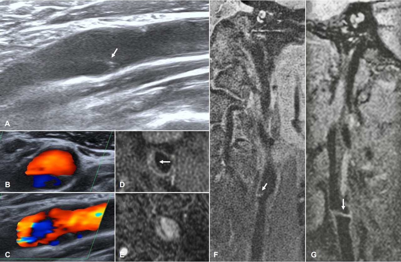
50세 남자가 건강검진으로 시행한 경동맥초음파에서 관찰된 죽경화판 소견으로 내원하였다. 경동맥초음파검사 이후에 아스피린, 항고혈압제, 항고지혈증제를 복용하고 있었으며, 15갑년의 흡연자였다. 추적검사상 이전 검사와 큰 차이는 없었으나 경동맥박리가 의심되어서(Fig. A-C) 정확한 진단을 위해 자기공명영상을 시행하였으며 경동맥갈퀴막(carotid web)으로 진단되었다(Fig. D-G).
경동맥갈퀴막은 경동맥망울에서 내강안쪽으로 튀어나온 선반 모양의 충만결손으로, 허혈뇌졸중의 위험인자로 연구되고 있다[1]. 경동맥갈퀴막을 검사하는 방법으로는 컴퓨터단층혈관조영검사 또는 고식적혈관조영술이 정확하지만 자기공명영상을 이용한 두개강 내외 혈관벽을 세밀하게 볼 수 있는 혈관벽영상(vessel wall imaging)을 이용하여 갈퀴막의 형태뿐만 아니라 혈관벽의 구성, 역학 및 혈류에 대한 중요한 정보를 얻을 수 있다[2].
경동맥갈퀴막은 조영증강자기공명영상에서 혈관벽의 두께 증가가 보이며, 혈관내강이 거짓분기(false bifurcation)로 나뉘는 것이 보인다. 이에 반해, 동맥박리에서는 유동신호의 소실(absent flow-void), 혈관 윤곽의 이상 소견(혈관벽의 불규칙성, 혈관내강의 협착 소견, 가성동맥류 등)이 관찰될 수 있다.

REFERENCES

1. Sajedi PI, Gonzalez JN, Cronin CA, Kouo T, Steven A, Zhuo J, et al. Carotid bulb webs as a cause of "cryptogenic" ischemic stroke. AJNR Am J Neuroradiol 2017;38:1399-1404.
crossref pmid
2. Mossa-Basha M, Alexander M, Gaddikeri S, Yuan C, Gandhi D. Vessel wall imaging for intracranial vascular disease evaluation. J Neurointerv Surg 2016;8:1154-1159.
crossref pmid pmc

Figure.
Ultrasonographic images and magnetic resonance images of the internal carotid artery. (A) Carotid ultrasonography shows a suspicious dissection flap (arrow) in the left proximal internal carotid artery. (B, C) Color Doppler ultrasonographic image shows turbulent flow. (D, E) T1-weighted axial and time-of-flight source magnetic resonance images show thin intramural protruding band-like lesion (arrow) arises from posteromedial wall of carotid bulb. Coronal (F) and sagittal (G) proton density-weighted magnetic resonance images show the carotid web clearly (arrow), in which no definitively visible intramural hematoma is documented.
jkna-36-3-260f1.jpg


ABOUT
BROWSE ARTICLES
EDITORIAL POLICY
FOR CONTRIBUTORS
Editorial Office
(ZIP 03163) #1111, Daeil Bldg, 12, Insadong-gil, Jongno-gu, Seoul, Korea
Tel: +82-2-737-6530    Fax: +82-2-737-6531    E-mail: jkna@neuro.or.kr                

Copyright © 2024 by Korean Neurological Association.

Developed in M2PI

Close layer