

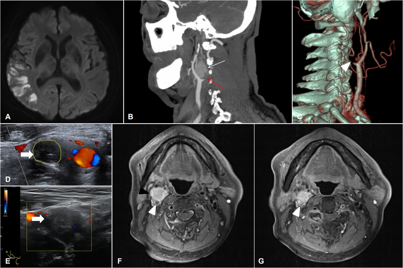
기저질환이 없는 68세 남자 환자가 좌측 위약감으로 내원하여 시행한 검사에서 중대뇌동맥영역에 급성뇌경색이 확인되었다(Fig. A). CT혈관조영술에서 우측 경동맥폐색 이외에 경동맥 분지부 주변에 조영증강되는 병변이 보였고(Fig. B, C), 경동맥초음파에서 우측 경동맥 혈류가 보이지 않고 그 주변으로 저에코병변이 관찰되었다(Fig. D, E). 두경부의 경동맥 분지부위에 호발하는 부신경절종(paraganglioma)이 의심되어 경부 MRI로 확인하였다(Fig. F, G). 종양 제거 수술을 하였고 병리 소견에서도 부신경절종이 확인되었다. 부신경절종은 신경릉세포(neural crest cells)에서 기원하는 종양으로[1], 경동맥 분지부와 목정맥구멍 그리고 미주신경에서 호발한다[2]. 본 환자는 종양의 침투가 경동맥폐색을 유발하면서 급성뇌경색이 발생하여 부신경절종을 진단한 드문 경우로 보고하였다.
| J Korean Neurol Assoc > Volume 36(3); 2018 > Article |

|
Figure.
Brain magnetic resonance image (MRI) shows acute right middle cerebral artery territorial infarction at the diffusion weighted sequence (A). Brain CT angiography show proximal internal carotid artery (ICA) occlusion (red arrow, B) (white arrow head, C) and mass at carotid bifurcation (white arrow, B). Carotid color doppler sonography reveal absent flow in proximal ICA (white arrow, D). Hypoechoic mass is detect in carotid sonography (white arrow, E). Mass (black arrow head) and ICA wall (white arrow head) are enhanced on the axial post-gadolinium T1-weighted MRI (F). Mass encase cervical ICA and similar signal intensity is shown in ICA lumen (white arrow head, G). CT; computed tomography.
- TOOLS
 PDF Links
 PubReader
 ePub Link
 Full text via DOI
 Download Citation
 Print
Print


-
METRICS

-
- 0 Crossref
- 0 Scopus
- 3,888 View
- 111 Download
-

- Related articles
- Editorial Office
-
(ZIP 03163) #1111, Daeil Bldg, 12, Insadong-gil, Jongno-gu, Seoul, Korea
Tel: +82-2-737-6530 Fax: +82-2-737-6531 E-mail: jkna@neuro.or.kr
Copyright © 2024 by Korean Neurological Association.

