경막하출혈로 이어진 자발두개내압 저하

Spontaneous Intracranial Hypotension Followed by Subdural Hemorrhage

Article information

J Korean Neurol Assoc. 2018;36(1):59-60
Publication date (electronic) : January 31, 2018
doi : http://dx.doi.org/10.17340/jkna.2018.1.16
Department of Neurology, Inha University Hospital, Inha University School of Medicine, Incheon, Korea
aDepartment of Neurology, Seoul Paik Hospital, Inje University College of Medicine, Seoul, Korea
황혜원, 윤병남,a
인하대학교 의과대학 부속병원 신경과
a인제대학교 의과대학 서울백병원 신경과
Address for correspondence: Byung-Nam Yoon, MD Department of Neurology, Seoul Paik Hospital, Inje University College of Medicine, 9 Mareunnae-ro, Jung-gu, Seoul 04551, Korea Tel: +82-2-2270-0045 Fax: +82-2-2270-0045 E-mail: nrybn1230@gmail.com
received : February 1, 2017 , rev-recd : November 24, 2017 , accepted : November 24, 2017 .

38세 여자가 수일 전부터 시작된 기립두통과 구토, 이명으로 내원하였다. 경미한 경부강직이 있었고, 감염증상은 없어 자발두개내압 저하(spontaneous intracranial hypotension)가 의심되었으나 치료를 거부하고 지내다가, 1개월 후 기립두통이 악화되어 재내원하였다. 뇌 자기공명영상에서 양측 뇌경막하삼출(subdural effusion)이 있었으며 경수막(pachymeninges)의 균등한 조영증강, 소뇌편도 탈출이 있어 두개내압 저하에 합당한 소견이었다(Fig. A-D) [1]. 요추천자를 하였으나 뇌척수액 채취는 불가하였다(dry tap). 침상안정, 정맥내 수액 등의 보존 치료와 경막외혈액첩포술(epidural blood patch)을 시행한 후 증상이 호전되어 퇴원하였고, 며칠 뒤 증상이 재발하였음에도 진료하지 않고 지내오다 1개월 후 의식혼탁과 구음장애로 응급실을 내원하였다. 뇌 전산화단층촬영에서 경막하출혈(subdural hemorrhage)이 발견되어 수술적 배액을 시행하였다(Fig. E, F) [2]. 자발두개내압 저하는 재발이 잦고, 악화될 경우 경막하출혈 등 심각한 신경손상의 위험이 있어 항상 적극적인 진단과 치료가 필요하다.
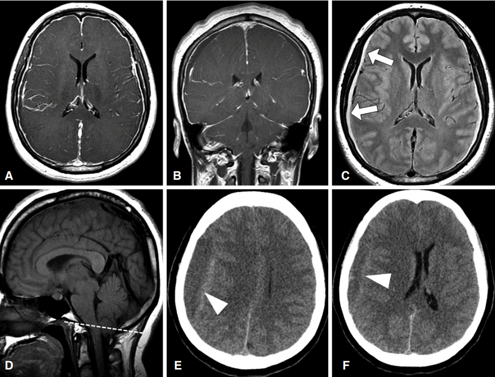
Figure.

Initial brain magnetic resonance imaging and follow-up brain CT. Brain T1-gadolinium-enhanced axial (A) and coronal (B) images show diffuse pachymeningeal enhancement; Axial T2-weighted FLAIR image (C) shows bilateral subdural fluid collection (arrows); Sagittal T1-weighted image (D) shows descent of the cerebellar tonsil through foramen magnum (dotted line); Brain CT (E, F) shows a right-sided subdural hemorrhage (arrowheads) with midline shift which causes reduction in the conscious level and dysarthria. CT; computed tomography, FLAIR; fluid attenuated inversion recovery.

References

1. Paldino M, Mogilner AY, Tenner MS. Intracranial hypotension syndrome: a comprehensive review. Neurosurg Focus 2003;15:ECP2.
2. de Noronha RJ, Sharrack B, Hadjivassiliou M, Romanowski CA. Subdural haematoma: a potentially serious consequence of spontaneous intracranial hypotension. J Neurol Neurosurg Psychiatry 2003;74:752–755.

Article information Continued

Figure.

Initial brain magnetic resonance imaging and follow-up brain CT. Brain T1-gadolinium-enhanced axial (A) and coronal (B) images show diffuse pachymeningeal enhancement; Axial T2-weighted FLAIR image (C) shows bilateral subdural fluid collection (arrows); Sagittal T1-weighted image (D) shows descent of the cerebellar tonsil through foramen magnum (dotted line); Brain CT (E, F) shows a right-sided subdural hemorrhage (arrowheads) with midline shift which causes reduction in the conscious level and dysarthria. CT; computed tomography, FLAIR; fluid attenuated inversion recovery.