분만 후 여성에서 발생한 거대동맥류에 의한 경동맥해면정맥동루
Carotid-Cavernous Fistula Due to Giant Aneurysm in a Postpartum Woman
Article information
Trans Abstract
A carotid-cavernous fistula (CCF) is an abnormal communication between the venous cavernous sinus and the carotid artery. The rupture of an intracavernous aneurysm is usually caused by trauma, but spontaneous rupture can also occur, with pregnancy being a contributing factor. We report a case of direct CCF due to rupture of a giant aneurysm in a postpartum woman.
경동맥해면정맥동루(carotid-cavernous fistula, CCF)는 외상이나 특발성으로 경동맥계와 해면정맥동 사이에 단락(shunt)이 형성되어 비정상적 교통이 일어난 상태를 말한다[1]. CCF의 증상은 누공의 크기와 위치에 따라 다양하지만 특징적으로 편측의 안구돌출, 충혈, 결막부종, 눈꺼풀처짐 및 외안근마비 등이 복합적으로 나타난다[2]. 원인에 따라 외상성과 자발성으로 나눌 수 있고, 혈류 흐름에 따라 고혈류형과 저혈류형으로 분류하기도 하며, 해부학적 위치에 따라 직접형과 간접형으로 구분할 수 있다[3].
내경동맥의 동맥류 파열은 자발성 CCF의 드문 원인으로 알려져 있다. 내경동맥 동맥류는 점차 크기가 증가하여 신경학적 증상을 나타내며, 파열될 경우 거미막하출혈보다는 CCF를 일으키는 경우가 더 흔하다[4]. 분만 후 여성에서 동맥류는 드물게 발생하나, 임신 중 호르몬과 혈역학적 변화가 새로운 동맥류의 형성과 기존 동맥류의 악화를 초래할 수 있는 것으로 알려져 있다[5].
저자들은 분만 후 여성에서 거대동맥류 파열에 의해 발생한 CCF를 경험하였으며, 코일 색전술을 통해 완전한 증상 회복을 보였기에 보고하고자 한다.
증 례
34세 여성이 3주간의 우측 안와와 전두부위의 통증과 결막충혈로 내원하였다. 두통은 우 편측에 발생하였으며 박동성 양상으로 내원 3일 전부터는 구토를 동반하였다. 과거력상 전신 질환의 병력 및 외상력은 없었으나 내원 2주 전 자연분만으로 남아를 출산한 상태였다. 입원 당시 활력징후는 혈압 130/90 mmHg, 체온 36.6℃, 맥박수 61회/분, 호흡수 20회/분이었다. 진찰에서 우측 안와의 부종, 결막 충혈 및 안구 부위 잡음(orbital bruit)이 보였다. 안과검사에서 시력은 정상이었으나 우안의 안구돌출이 있었고, 우안의 안압(18 mmHg)이 좌안(9 mmHg)보다 증가되어 있었다. 신경학적 진찰에서 우안의 외전운동 제한이 있었지만 다른 이상소견은 없었다.
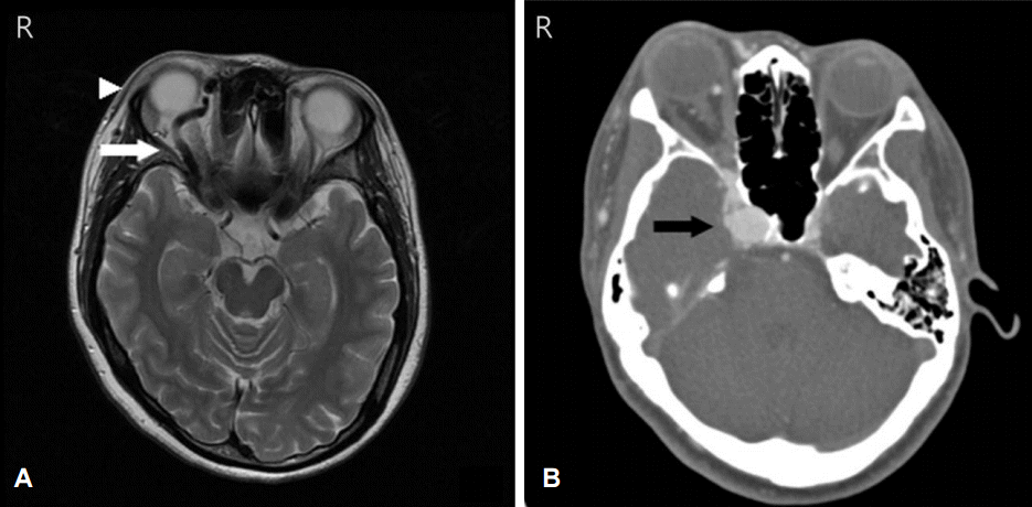
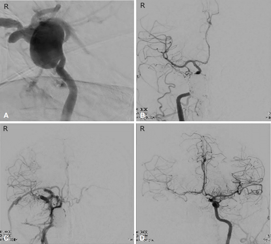
응급으로 뇌MRI와 CT혈관조영술을 한 결과 우측 경동맥의 거대 동맥류(3.2×1.4 cm) 파열로 인한 CCF를 확인하였다(Fig. 1). 오른쪽 대퇴동맥을 천자하여 오른쪽 내경동맥에 유도 도관을 설치한 후 미세철사를 이용하여 미세도관을 동맥류 내부에 설치하였다. 동맥류 입구에 미세도관을 하나 더 설치하였고, 스텐트(CODMAN ENTERPRISE®, Codman & Shurtleff Inc., Raynham, MA, USA) 도움으로 코일링을 시도하였다. 우측 경동맥을 혈관조영하여 동맥류가 완벽하게 색전된 것을 확인하였고 우측 CCF는 폐쇄되었다(Fig. 2A, B). 시술 다음 날부터 우측 안와 부종과 안구부위 잡음은 크게 호전되었다.

Brain MRI and CT angiography on admission. (A) Axial T2-weighted MRI shows engorged right superior ophthalmic vein (arrow) and proptosis of right eye (arrowhead). (B) CT angiography demonstrates a giant aneurysm and flow into the cavernous sinus during the early arterial phase (arrow). MRI; magnetic resonance imaging, CT; computerized tomography.

Transfemoral angiography (A) Transfemoral angiography on admission shows CCF with rupture of large aneurysm (3.2×1.4 cm) in the cavernous segment of right ICA. (B) The CCF and aneurysm were completely occluded after coil embolization. (C) Transfemoral angiography after aggravated symptoms showed a recurred CCF. (D) Right ICA was completely obliterated and collateral flow through left ICA was confirmed. CCF; carotid-cavernous fistula, ICA; internal carotid artery.
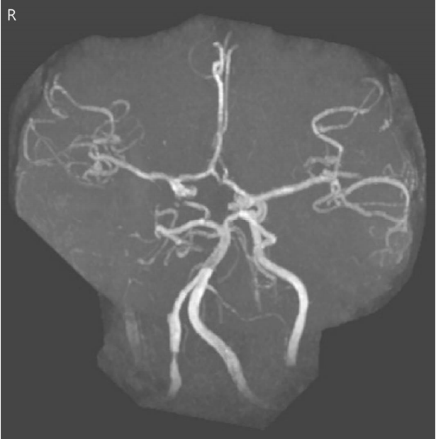
그러나 시술 4일째 되는 날, 환자의 두통이 악화되었고 안와 부종과 안구부위 잡음이 다시 발생하였다. 뇌혈관조영술에서는 CCF의 재발이 확인되었다(Fig. 2C). 동맥류의 코일링을 다시 시도하였으나 CCF가 지속적으로 확인되어 우측 내경동맥의 폐쇄를 계획하였다. 좌측 대퇴동맥도 천자하여 이중 도관을 이용하였으며, 좌측 내경동맥과 앞교통동맥을 통해 우측 원부위 내경동맥에 도달하였다. 곁순환을 확인한 뒤 코일색전술을 통해 우측 내경동맥을 완전 폐쇄하였고 CCF 역시 완전히 폐쇄된 것을 확인하였다(Fig. 2D). 두 번째 시술 이후, 환자는 약간의 두통만 있었고, 결막충혈, 안와부종 및 우안의 외전장애는 모두 호전되었다. 추적 MR혈관조영술에서도 CCF의 재발은 없었다(Fig. 3). 시술 2주일째 환자는 퇴원하였고, 항혈소판제로 아스피린을 복용중이며 퇴원 후 7개월까지 특별한 합병증 없이 다음 임신을 계획하고 있다.
고 찰
Barrow 등[3]은 병리학, 혈역학 및 혈관조영술 상의 특징에 따라 CCF의 분류를 제시하였는데 두부외상이나 동맥류의 파열에 의해 내경동맥과 해면정맥동 사이의 직접 단락이 발생하는 경우는 A형, 자연적으로 발생하는 경막 CCF 중 내경동맥의 뇌막분지와 연결되는 경우는 B형, 외경동맥의 뇌막분지와 연결되는 경우는 C형, 내경동맥과 외경동맥 양쪽의 뇌막분지와 연결되는 경우는 D형으로 구분하였다. A형은 빠른 혈류에 의해 특징적인 증상이 저명하게 나타나며, B-D형은 뇌막혈관을 통한 느린 혈류에 의해 비전형적인 눈 운동장애 소견만 나타날 수도 있다. 저자들의 증례는 해면정맥동에 위치한 거대동맥류 파열로 인해 CCF가 발생한 경우이므로 A형에 해당한다.
CCF는 해면정맥동 정맥흐름의 방향에 따라 임상증상의 차이를 보일 수 있다. 방향은 전방, 후방, 전후방으로 나눌 수 있는데, 위눈정맥(superior ophthalmic vein)을 포함하는 전방 누공으로 배출되는 경우 주로 안와-안구 울혈증상을 보이고(red-eyed shunt), 후방 누공으로 배출되는 경우 안구운동마비와 두통을 주 증상으로 하는 백색안구션트(white-eye shunt) 증상이 나타날 수 있다[2]. 이외에도 뇌막분지(cortical vein)로 배출되는 경우 정맥압을 상승시켜 두개강내출혈을 유발할 수 있고, 동맥류가 경막을 침범하여 거미막하공간에 걸쳐 위치하는 경우 거미막하출혈을 일으킬 수 있다[6]. 증례의 환자는 뇌영상검사와 혈관조영술을 통해 위눈정맥으로 정맥흐름을 확인하였고, 따라서 안와-안구 울혈 증상을 보이는 경우에 해당한다. 주요 뇌막분지로의 정맥 흐름은 확인되지 않았기 때문에 두개강내출혈이 동반되지 않았던 것으로 생각된다.
해면정맥동 분절에 위치한 내경동맥류는 전체 뇌동맥류의 약 2-5%, 내경동맥에 생기는 동맥류 중 10-15%를 차지하며 주로 50대 이상의 여성에서 잘 생기는 것으로 보고 되어있다[7]. van Rooij 등6 에 따르면 뇌동맥류중 CCF를 일으키는 경우는 전체 689예 중 10예(1.5%)이며, 혈관내 중재시술을 통해 확인된 증상이 있는 파열 뇌동맥류 41예 중 10예(24.4%)를 차지하였다. 저자들의 환자와 같이 거대 내경동맥류(3.2×1.4 cm) 파열로 CCF가 발생한 경우는 매우 드물다.
CCF의 다수는 외상성으로 발생하나 자발성인 경우도 적지 않게 보고 되어 있고 특히 여성은 남성보다 자발성 CCF 발생위험이 높은 것으로 알려져 있다[8]. 임신은 자발성 CCF의 주요 발생 원인이며, 환자 중 25-30%는 임신 후반기 혹은 출산 중에 CCF가 발생한 것으로 보고되어 있다[9]. 임신 후반기에는 정맥압이 증가하고 혈관에 대한 에스트로겐 효과가 극대화된다. 이러한 변화는 고혈압을 유발하여 새로운 동맥류의 형성을 증가시키고 이전에 있던 동맥류의 악화를 초래할 수 있다[5]. 저자들의 환자는 이전에 뇌영상검사를 한 적이 없어 동맥류가 기존에 있었는지는 알 수 없으나, 특별한 과거력이 없었던 점을 고려할 때 임신과 출산이 거대동맥류의 발생에 기여하였을 것으로 추정할 수 있다.
저자들은 외상의 병력이 없는 분만후 여성에서 CCF와 동반된 거대동맥류를 확인하였고 코일색전술을 통해 증상을 완전히 치료한 증례를 경험하여 보고하는 바이다.
