

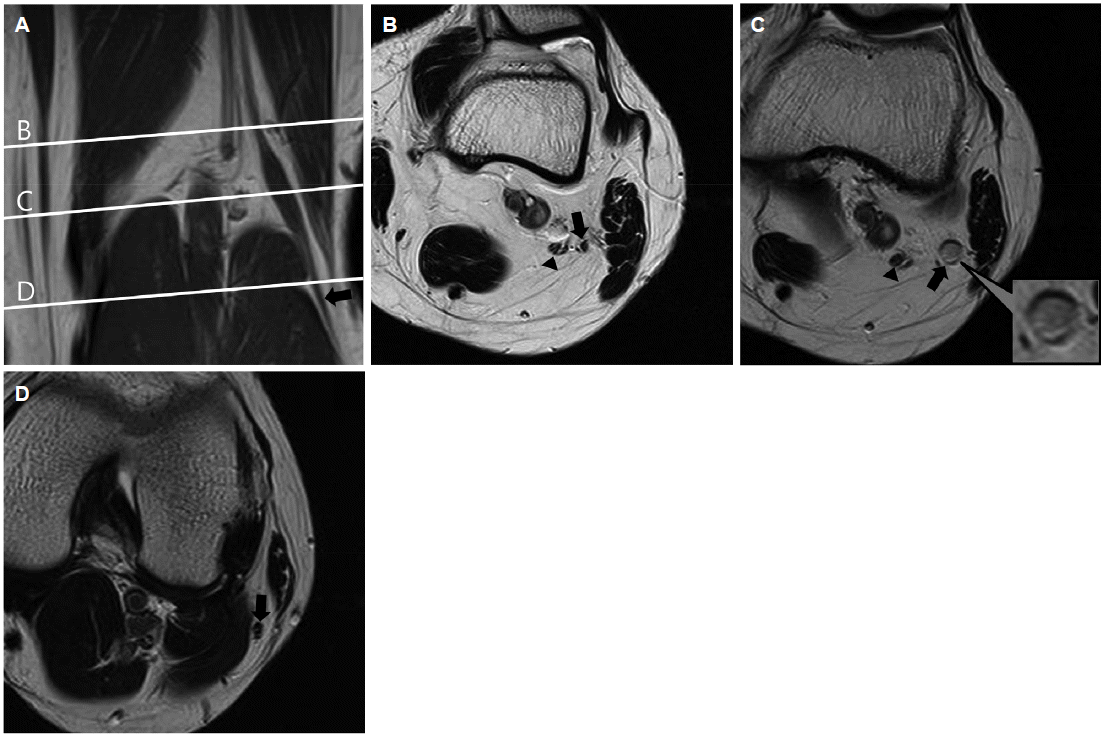
외상의 병력 없이 20세 여자가 왼쪽 발처짐으로 왔다. 발처짐은 2년 전 발생하여 점차 심해졌고, 발가락위약도 발생하였다. 신경학적 진찰에서 왼쪽 발등굽힘과 외번의 위약이 있었고 발바닥쪽굽힘과 내번은 정상이었다. 신경전도검사와 근전도검사에서 왼쪽 종아리신경마비로 판단하였으며 Philips iU22 고해상도 초음파기계(Philips Healthcare, Eindhoven, The Netherlands)의 12-MHz 선형탐색자를 이용한 초음파에서 대퇴골관절융기주위의 종아리신경 안에서 30 mm × 8 mm × 5 mm 크기의 저에코의 가늘고 긴 종괴가 관찰되었다(Fig. 1). 이후 촬영한 무릎 자기공명영상에서 같은 부위에 신경성종양으로 확인되었다(Fig. 2). 종아리신경마비 환자의 7.6%에서 종양이 관찰된다[1]. 단일신경병의 명확한 원인이 없을 때 신경초음파는 병변의 위치뿐만 아니라 신경비후나 구조적이상에 대한 정보를 제공하여 진단적 가치가 있다[2].
| J Korean Neurol Assoc > Volume 33(3); 2015 > Article |
REFERENCES
1. Kim DH, Murovic JA, Tiel RL, Kline DG. Management and outcomes in 318 operative common peroneal nerve lesions at the Louisiana State University Health Sciences Center. Neurosurgery 2004;54:1421-1428.


2. Visser LH, Hens V, Soethout M, De Deugd-Maria V, Pijnenburg J, Brekelmans GJ. Diagnostic value of high-resolution sonography in common fibular neuropathy at the fibular head. Muscle Nerve 2013;48:171-178.
Figure 1.
Ultrasonogram. Transverse view of left common peroneal nerve (white arrow) shows hypoechogenecity and enlargement (A). Longitudinal view shows normal proximal common peroneal nerve (arrowheads) which is enlarged (white arrows) in the femoral condylar level (B). TN; posterior tibial nerve.
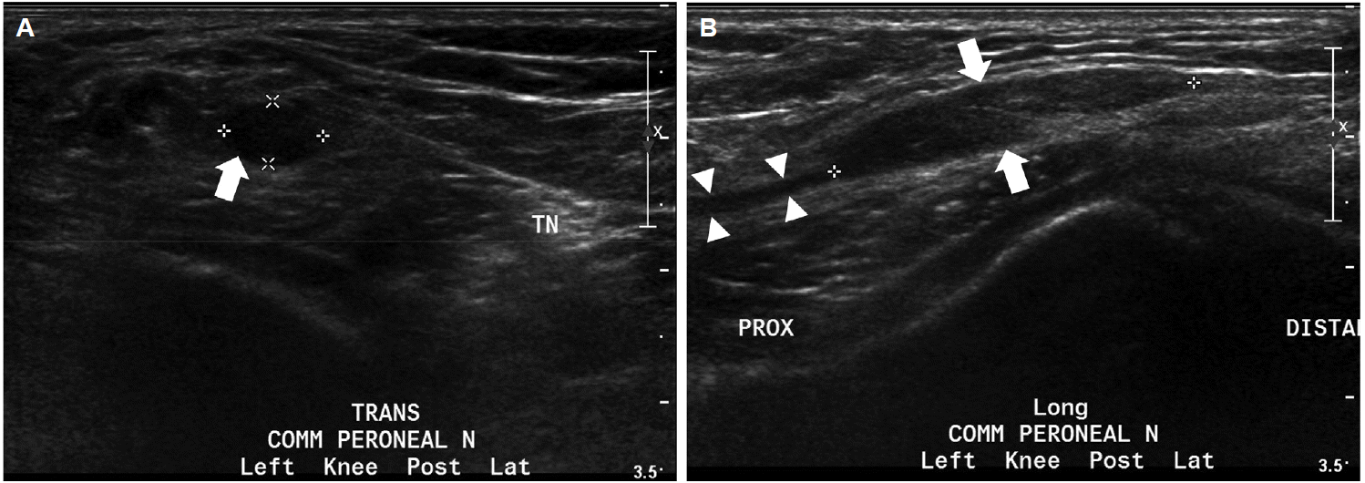
Figure 2.
Knee MRI. Coronal T1-weighted (A) and transverse T2-weighted images (B, C, D) showing left posterior tibial nerve (arrowheads) and common peroneal nerve (black arrows). A mass is noted near the femoral condyle (A, C). Proximal (B) and distal (D) to the mass is normal common peroneal nerve (B, D). MRI; magnetic resonance imaging.
- TOOLS
 PDF Links
 PubReader
 ePub Link
 Full text via DOI
 Download Citation
 Print
Print


-
METRICS

-
- 0 Crossref
- 0 Scopus
- 5,991 View
- 120 Download
-

- Related articles
- Editorial Office
-
(ZIP 03163) #1111, Daeil Bldg, 12, Insadong-gil, Jongno-gu, Seoul, Korea
Tel: +82-2-737-6530 Fax: +82-2-737-6531 E-mail: jkna@neuro.or.kr
Copyright © 2024 by Korean Neurological Association.

