아급성연합변성의 자기공명영상 소견
Magnetic Resonance Imaging in Subacute Combined degeneration
Article information
71세 여자가 2개월 전부터 시작된 보행장애와 최근에 발생한 양측 상하지의 감각저하를 주 증상으로 병원에 왔다. 19년 전 당뇨로 진단받고 약물 복용 중이었고, 11년 전 조기위암으로 대부분위절제술을 받았다. 신경학적진찰에서 감각실조 외에 하지의 고유감각저하가 관찰되어, 아급성연합변성으로 진단하고 비타민 B12를 근육주사하였다. 근육주사 전 시행한 비타민 B12는 81 pg/mL로 저하되어 있었다. 척추 시상면 자기공명영상에서 경수 및 흉수의 뒤기둥을 따라 T2강조영상에서 고신호강도가 관찰되었다. 축방향 T2강조영상 및 확산강조영상에서는 뒤기둥 이외에도 가쪽척수시상로에서도 고신호강도가 확인되었다(Fig. A,B). 해당 병변은 겉보기확산계수지도에서 고신호강도를 보여(Fig. C,D) 세포독성뇌부종을 동반한 Waller변성으로 판단하였다. 현재까지 아급성연합변성의 임상소견은 잘 알려져 있으나[1,2], 확산강조영상을 포함한 자기공명영상 소견은 국내보고가 없어 보고하고자 한다.
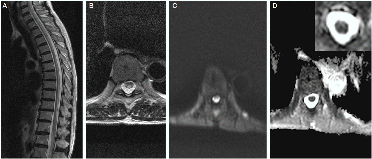
MR image. T2 weighted sagittal (A) and axial (B) image showed a long extened high signal intensity lesion along the posterior column and the lateral spinothalamic tract. Diffusion (C) image showed a high signal intensity lesion, and apparent diffusion coefficient image showed a low signal intensity lesion at the corresponding area.