낮은 전압에 의한 전기화상 환자에서 발생한 척수병증
Myelopathy in a Patient with Low-Voltage Electrical Burn
Article information
J Korean Neurol Assoc. 2015;33(1):66-66
Publication date (electronic) : February 1, 2015
doi :
http://dx.doi.org/10.17340/jkna.2015.1.18
received : June 16, 2014 , rev-recd : July 3, 2014 , accepted : July 3, 2014 .
Keywords: Myelopathy electrical burn spinal cord
31세 남자가 6일 전부터 발생한 좌측 손의 근력 저하와 감각 이상으로 병원에 왔다. 증상은 7일 전 300볼트 전압의 제분기에 감전된 다음날부터 발생하였다. 신경학적 진찰에서 좌측 손의 쥐는 힘이 MRC (medical research council) 2등급으로 저하되어 있었으며 손목 굽힘, 폄 운동은 4등급 정도였다. 척수자기공명영상에서 C1부터 T4 영역까지 출혈을 동반한 척수병증이 관찰되었다(Fig. ). 전기에 의한 신경계 손상은 본 증례처럼 척수 손상의 형태로 나타날 수 있다. 잠복기는 다른 신경계 손상에 비해 늦게 출현한다고 알려져 있으나 본 증례처럼 1일 이내에 나타난 경우도 드물지 않다[1]. 척수자기공명영상에서 이상이 나오는 경우는 적고 본 증례처럼 1,000 볼트 이하의 낮은 전압에 의해 발생한 척수병증에 대한 보고는 매우 드문 것으로 알려져 있다[1,2].
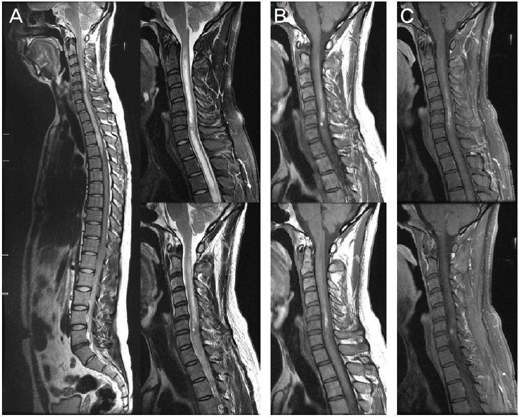
Spinal MRI. (A) There are increased T2-weighted signal intensities of spinal cord at C1-T4 (B) T1-weighted MRI shows the hyperintense lesion at C2-T1, suggesting myelopathy with hemorrhage. (C) Hemorrhagic lesions are enhanced on Gadolinium enhanced T1-weighted MRI.
References
1. Nam KS, Cha MJ, Kim MJ, Oh MS, Minn YK, Cho SJ, et al. Features of the myelopathy in patients with electrical burn. J Korean Neurol Assoc 2007;25:180–186.
2. Jha S, Singh MN. Acute transverse myelitis following electrical injury: a short report. Neurol India 2001;49:321–322.
Article information Continued
Copyright © 2015 Korean Neurological Association