

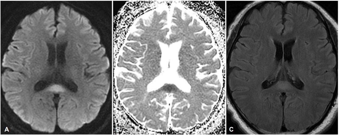
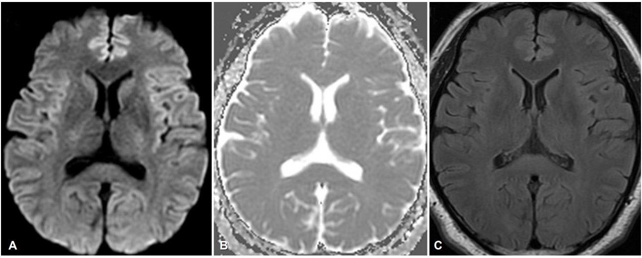
기저질환이 없는 43세 남자가 글루포시네이트암모늄이 함유된 제초제를 마신 후 발생한 의식 혼미로 응급실에 왔다. 입원 후 2-3일에 의식 혼미가 지속되었고, 수차례 전신강직간대발작이 있었다. 입원 4일째 시행한 뇌 자기공명영상 확산강조영상과 액체감쇠역전회복영상에서 뇌량팽대에 저명한 고신호강도 병변이 보였다(Fig. 1). 이 병변은 조영증강되지 않았다. 뇌파검사에서는 지속서파가 전반적으로 있었다. 환자는 투석 및 수액 공급 등 내과적 치료 후 회복하여 퇴원하였다. 4달 뒤 추적 뇌 자기공명영상에서 이전에 보였던 병변은 사라졌다(Fig. 2).
| J Korean Neurol Assoc > Volume 37(3); 2019 > Article |

|
REFERENCES
1. Kim JH, Yu I, Kim YD, Na SJ, Lee KO, Yoon B. Encephalopathy after glufosinate ammonium intoxication. J Korean Neurol Assoc 2014;32:113-116.
2. Jeong TO, Yoon JC, Lee JB, Jin YH, Hwang SB. Reversible splenial lesion syndrome (RESLES) following glufosinate ammonium poisoning. J Neuroimaging 2015;25:1050-1052.


- TOOLS
 PDF Links
 PubReader
 ePub Link
 Full text via DOI
 Download Citation
 Print
Print


-
METRICS

-
- 0 Crossref
- 0 Scopus
- 2,932 View
- 58 Download
-

- Related articles
-
Reversible Lesion in Splenium of Corpus Callosum Following COVID-19 Vaccination2022 May;40(2)
Reversible Cerebral Vasoconstriction Syndrome Associated with Severe Anemia2018 August;36(3)
Reversible Parkinsonism Associated with Acute Arsenic Intoxication2011 ;29(3)
- Editorial Office
-
(ZIP 03163) #1111, Daeil Bldg, 12, Insadong-gil, Jongno-gu, Seoul, Korea
Tel: +82-2-737-6530 Fax: +82-2-737-6531 E-mail: jkna@neuro.or.kr
Copyright © 2024 by Korean Neurological Association.

