

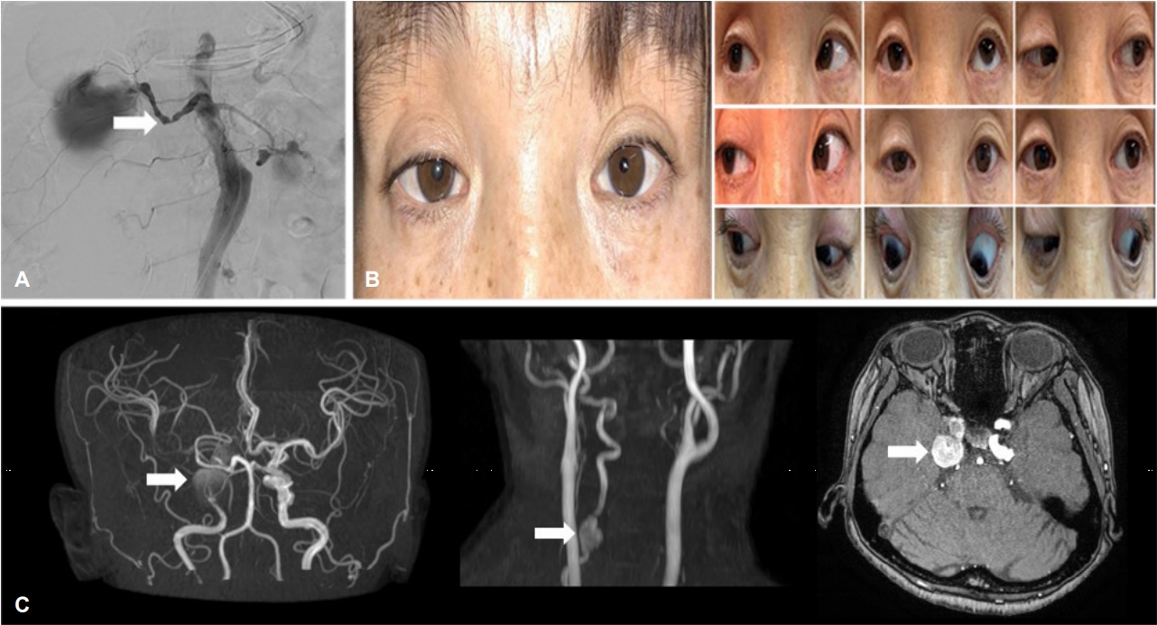
45세 여자가 일주일 전에 발생한 어지럼과 두통으로 왔다. 환자는 섬유근형성이상(fibromuscular dysplasia)으로 진단받은 후 우측 콩팥동맥 협착으로 피부경유혈관경유관풍선혈관성형술(percutaneous transluminal balloon angioplasty)을 시행받은 과거력이 있었다(Fig. A). 신경학적 진찰에서 우측 눈 안검하수와 우측 눈의 동공이 좌측에 비해 확장되어 있는 동공부등(anisocoria) 및 동공반사가 우측에서 느리게 관찰되었다(Fig. B). 안구운동검사에서는 우측 눈의 상방주시가 불가능한 상태였다(Fig. B). 뇌 자기공명혈관조영 영상에서 우측 경동맥에 2 cm 크기의 뇌동맥류와 함께 좌측 경동맥, 척추동맥에서도 뇌동맥류가 확인되었다(Fig. C).
| J Korean Neurol Assoc > Volume 37(2); 2019 > Article |

|
REFERENCES
1. Lee DK, Heo SH, Kwon SH, Park KC, Ahn TB, Yoon SS, et al. Cerebral infarction in a young female patient with renovascular hypertension caused by fibromuscular dysplasia. J Korean Neurol Assoc 2010;28:326-328.

2. Bhatti MT, Eisenschenk S, Roper SN, Guy JR. Superior divisional third cranial nerve paresis: clinical and anatomical observations of 2 unique cases. Arch Neurol 2006;63:771-776.

Figure.
(A) Renal artery angiography showed stenotic lesion (string of beads sign) of the right renal artery (arrow). (B) Note the right upper eyelid ptosis and pupil dilatation. Nine positions of gaze. There is limitation of elevation of the right eye. (C) MRA showed aneurysms in the intra-cavernous part of both internal carotid artery and right vertebral artery (arrow). Axial T1-enhanced image showed aneurysms on cavernous sinus (arrow). MRA; magnetic resonance angiography.
- TOOLS
 PDF Links
 PubReader
 ePub Link
 Full text via DOI
 Download Citation
 Print
Print


-
METRICS

-
- 0 Crossref
- 0 Scopus
- 3,331 View
- 79 Download
-

- Related articles
-
Isolated Hypoglossal Nerve Palsy Caused by Dural Arteriovenous Fistula2016 November;34(4)
Isolated Bilateral Abducens Nerve Palsy Caused by Basilar Artery Dissecting Aneurysm2012 ;30(4)
Two Cases of Oculomotor Nerve Palsy Due to Dural Carotid ?Cavernous Fistula 1995 ;13(3)
- Editorial Office
-
(ZIP 03163) #1111, Daeil Bldg, 12, Insadong-gil, Jongno-gu, Seoul, Korea
Tel: +82-2-737-6530 Fax: +82-2-737-6531 E-mail: jkna@neuro.or.kr
Copyright © 2024 by Korean Neurological Association.

